This human anatomy ClipArt gallery offers 140 illustrations of the peripheral nervous system, which includes the nervous structures not included in the central nervous system such as peripheral nerves that extend from the spine through the torso and limbs.

Diagram showing relation of neurons composing the spinal nerve roots with adjacent nervous structures. Labels: S.E., sensory epithelium connected by a sensory neuron with spinal cord; S.M., striated muscle receiving the axon from a motor-cell in the ventral horn of the gray matter in the cord; Sp. F., spinal fibers, medullated, sensory, and the motor, passing to the sympathetic ganglion where they connect with sympathetic neurons; S.F., S.F., non-medullated fibers from the sympathetic neurons passing to the viscera, the axons going to the plain muscle (P.M.), the dendrites to the sensory endings (S.E.).

Spinal Nerve Roots

Diagram showing relation of neurons composing the spinal nerve roots with adjacent nervous structures.…

Scheme of the distribution of a typical spinal nerve.

Distribution of a Spinal Nerves

Scheme of the distribution of a typical spinal nerve.

The brachial plexus of the spinal nerves.

The Brachial Plexus of the Spinal Nerves

The brachial plexus of the spinal nerves.

The brachial plexus of the spinal nerves, and nerves of the upper extremity.

The Brachial Plexus of the Spinal Nerves

The brachial plexus of the spinal nerves, and nerves of the upper extremity.

The cervical plexus of the spinal nerves.

The Cervical Plexus of the Spinal Nerves

The cervical plexus of the spinal nerves.

The lumbar and sacral plexuses of the spinal nerves.

The Lumbar and Sacral Plexuses of the Spinal Nerves

The lumbar and sacral plexuses of the spinal nerves.

The lumbar and sacral plexuses of the spinal nerves, showing the distribution of nerve branches.

The Lumbar and Sacral Plexuses of the Spinal Nerves

The lumbar and sacral plexuses of the spinal nerves, showing the distribution of nerve branches.
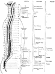
Table giving approximate areas of distribution of the different spinal nerves with a diagram showing their respective levels of exit from the vertebral column.

Distribution of the Spinal Nerves

Table giving approximate areas of distribution of the different spinal nerves with a diagram showing…
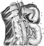
The spinal nerves, sympathetic cord, and the network of sympathetic nerves around the internal organ. Labels: K, aorta; A, esophagus; B, diaphragm; C, stomach.

Spinal Nerves, Sympathetic Cord, and the Network of Sympathetic Nerves

The spinal nerves, sympathetic cord, and the network of sympathetic nerves around the internal organ.…

The sympathetic ganglions and their connection with other nerves. Labels: A, The semilunar ganglion and solar plexus. D, The thoracic (chest) ganglions. E, The external and internal branched of the thoracic ganglions. G, H, The right and left coronary (heart) plexus. I, N, Q , The inferior, middle and superior cervical (neck) ganglions. 1, The renal plexus of nerves. 2, The lumbar (loin) ganglions. 3, Their internal branches. 4, Their external branches. 5, The aortic plexus of nerves.

The Sympathetic Ganglions and their Connection to other Nerves

The sympathetic ganglions and their connection with other nerves. Labels: A, The semilunar ganglion…
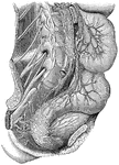
The great sympathetic nerve.

The Great Sympathetic Nerve

The great sympathetic nerve.

"Showing the distribution of some of the great plexuses of the sympathetic nerve in the lumbar and sacral regions." — Blaisedell, 1904

Plexuses of the Sympathetic Nerves

"Showing the distribution of some of the great plexuses of the sympathetic nerve in the lumbar and sacral…

General view of the sympathetic nervous. Labels: 1,2,3, cervical ganglia; 4, 1st thoracic ganglion; 5, 1st lumbar ganglion; 6, 7, sacral ganglion; 9, 9, cardiac nerves; 13, branch of pneumogastric nerve ending in semi-lunar ganglion; 14, epigastric plexus.

General View of the Sympathetic Nervous System

General view of the sympathetic nervous. Labels: 1,2,3, cervical ganglia; 4, 1st thoracic ganglion;…

The sympathetic nervous system.

The Sympathetic Nervous System

The sympathetic nervous system.

Diagrammatic view of the Sympathetic cord of the right side, showing its connections with the principal cerebrospinal nerves and the main prae-aortic plexuses. Cerebrospinal nerves. VI, a portion of the sixth cranial as it passes through the cavernous sinus, receiving two twigs from the carotid plexus of the sympathetic nerve; O, opthalmic ganglion connected by a twig with the carotid plexus; M, connection of the spheno-palatine ganglion by the Vidian nerve with the carotid plexus; C, cervical plexus; Br, brachial plexus; D 6, sixth intercostal nerve; D 12, twelfth; L 3, third lumbar nerve; S 5, fifth; Cr, anterior crural nerve winding round the subclavian artery. Sympathetic Cord. c, superior cervical ganglion; c', second or middle; c", inferior; from each of these ganglia cardiac nerves (all deep on this side) are seen descending to the cardiac plexus; d 1, placed immediately below the first dorsal sympathetic ganglion; d 6, is opposite he sixth; l 1, first lumbar ganglion. Preaortic and Visceral Plexuses. p, pharyngeal, and, lower down, laryngeal plexus; pl, post-pulmonary plexus spreading from the vagus on the back of the right bronchus; c a,on the aorta , the cardiac plexus, towards which , in addition to the cardiac nerves from the three cervical sympathetic ganglia, other branches are seen descending from the vagus and recurrent nerves; co, right or posterior and co1, left or an. coronary plexus; o, esophageal plexus in long meshes on the gullet; sp, great splanchnic nerve formed by branched from the fifth, sixth, seventh, eight, and ninth dorsal ganglia; +, small splanchnic from the ninth and tenth; ++, smallest or third splanchnic from the eleventh: the first and second of theses are shown joining the solar plexus, s o; the third descending to the renal plexus, r e; connecting branches between the solar plexus and the vagi are also represented; pn', above the place where the right vagus passes to the lower or posterior surface of the stomach; pn", the left distribution on the anterior side or upper surface of the cardiac portion of the organ: from the solar plexus large branches are seen surrounding the arteries of the coeliac axis, and descending from the solar and sup. mesenteric plexuses and from the lumbar ganglia; mi, the inf. mesenteric plexus surrounding the corresponding artery; hy, hypogastric plexus; pl, the right pelvic or inf. hypogastric plexuses; pl, the right pelvic plexus; from this the nerves descending are joined by those from the plexus on the sup. hemorrhoidal vessels, mi', by nerves from the third and fourth sacral spinal nerves, and there are thus formed the rectal, vesical, and other plexuses, which ramify upon the viscera, ,as towards ir, and v, the rectum and bladder.

Sympathetic System

Diagrammatic view of the Sympathetic cord of the right side, showing its connections with the principal…
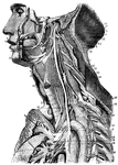
The sympathetic system of nerves.

The Sympathetic System of Nerves

The sympathetic system of nerves.

Diagram of the upper half of a male human showing the routes of the nervous system.

Human Nerve System

Diagram of the upper half of a male human showing the routes of the nervous system.

Nerves of the thigh. Labels: 1, gangliated cord of sympathetic; 2, third lumbar nerve; 3, branches to iliacus internus; 4, fourth lumbar nerve; 5, anterior crural nerve; 6, lumbosacral nerve; 7, branch to psoas; 8, obturator nerve; 9, external cutaneous nerve (cut); 10, nerve to pectineus; 11, superficial division of obturator nerve; 13, 13, sartorius muscle; 14, 14, adductor longus; 15, branch to rectus; 16, deep division of obturator nerve; 17, branches to vastus externus and crureus; 18, adductor brevis; 19, branch to vastus internus; 20, adductor magnus; 21, vastus externus; 22, 22, internal saphenous nerve; 23, rectus femoris; 24, pattelar branch of saphenous nerve; 25, vastus internus; 26, gracilis.

Nerves of the Thigh

Nerves of the thigh. Labels: 1, gangliated cord of sympathetic; 2, third lumbar nerve; 3, branches to…

Nerves of the thigh. Labels: 1, sympathetic ganglia; 2, third lumbar; 3, branches to iliacus; 4, fourth lumbar; 5, anterior crural; 6, lumbo-sacral; 7, branch of the psoas; 8, obturator; 9, external cutaneous; 10, nerve to pectineus; 11, branch anterior crural; 12, superficial division of obturator; 13, sartorius muscle; 14, adductor longus; 15, branch to rectus; 16, deep division of obturator; 17, branches to vastus externus and crureus; 18, adductor brevis; 19, branch to vastus internus; 20, adductor magnus; 21, vastus externus; 22, internal saphena; 23, rectus femorio; 24, patellar branch of saphena; 25, vastus internus; 26, gracilis.

Nerves of Thigh

Nerves of the thigh. Labels: 1, sympathetic ganglia; 2, third lumbar; 3, branches to iliacus; 4, fourth…
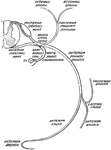
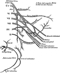
The 5th cranial nerve, also known as the trifacial or the trigeminus nerve.

Trifacial Nerve

The 5th cranial nerve, also known as the trifacial or the trigeminus nerve.