This human anatomy ClipArt gallery offers 73 illustrations of the human upper respiratory system, including organs involved in respiration. The human upper respiratory tract includes the nasal passages, pharynx, and the larynx.

Operative approaches through the front of the neck to the larynx, pharynx, and trachea. a: Approach to pharynx between the hyoid and the thyroid cartilages. b: Approach to the larynx between the thyroid and cricoid cartilages. c: approach to the larynx below the cricoid cartilage and above the isthmus of the thyroid gland. d: Approach for low tracheotomy.

Upper Respiratory Tract

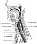
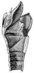
Operative approaches through the front of the neck to the larynx, pharynx, and trachea. a: Approach…

Diagram of rima glottidis. A, during ordinary easy breathing. B, widely opened.

Rima Glottidis

Diagram of rima glottidis. A, during ordinary easy breathing. B, widely opened.

Exposure of the right maxillary sinus, after removal of facial muscles. The (*) indicates the opening (Ostium maxillare) of the maxillary sinus into the nose.

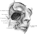
Dissection of the Maxillary Sinus

Exposure of the right maxillary sinus, after removal of facial muscles. The (*) indicates the opening…

Right pterygopalatine fossa, from without. The greater portion of the ala magna oss. sphenoid., of the lateral part of the corpus maxillae and of the pars orbit. oss. front. have been removed by a sagittal section, exposed the medial wall of the sinus maxillaris and of the orbit. Labels: **, indicate a probe through the Foramen rotundum; *, indicates a probe through the Canalis pterygoideus.

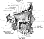
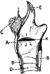
Pterygoid Fossa and Maxillary Sinus

Right pterygopalatine fossa, from without. The greater portion of the ala magna oss. sphenoid., of the…

Diagram of the pillars of the fauces and of the tonsils.

Tonsil and Pillars of the Fauces

Diagram of the pillars of the fauces and of the tonsils.

Tonsil of the nasopharynx of a boy aged 2 years.

Tonsil of the Nasopharynx

Tonsil of the nasopharynx of a boy aged 2 years.

"Larynx, Trachea, and the Bronchi. (Front view.) <em>A</em>, epiglottis; <em>B</em>, thyroid cartilage; <em>C</em>, cricothyroid membrane, connecting with the cricoid cartilage below, all forming the larynx; <em>D</em>, rings of the trachea." &mdash; Blaisedell, 1904

Trachea

"Larynx, Trachea, and the Bronchi. (Front view.) A, epiglottis; B, thyroid cartilage;…

Image of vocal apparatus as seen in the laryngoscopic mirror held far back in the mouth.

Vocal Apparatus

Image of vocal apparatus as seen in the laryngoscopic mirror held far back in the mouth.

"<em>A</em>, epiglottis; <em>B</em>, section of hyoid bone; <em>C</em>, false vocal cords which have nothing to do with the production of voice; <em>D</em>, oblong opening between the false and true vocal cords; <em>E</em>, true vocal cord;<em>F</em>, section of the thyroid cartilage; <em>H</em>, section of the anterior portion of the cricoid cartilage; <em>K</em>, trachea; <em>L</em>, section of the posterior portion of the cricoid cartilage; <em>M</em>, ladle-shaped cartilage." &mdash; Blaisedell, 1904

Vocal cords

"A, epiglottis; B, section of hyoid bone; C, false vocal cords which have…

This illustration shows the vocal cords as seen from above during phonation (A. Thyroid Cartilage; B. Cricoid Cartilage; C. hyoid bone; D. Rings of trachea; E. Epiglottis; F. Arytenoid Cartilage; G. Thyro-arytenoid muscle; H. Adam's Apple; I. False Vocal Cords; J. True Vocal Cords; K. Ventricle; L. Rima Glottis).

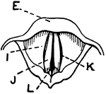
Vocal Cords Seen from above During Phonation

This illustration shows the vocal cords as seen from above during phonation (A. Thyroid Cartilage; B.…

This illustration shows the vocal cords, seen from above during quiet breathing (A. Thyroid Cartilage; B. Cricoid Cartilage; C. hyoid bone; D. Rings of trachea; E. Epiglottis; F. Arytenoid Cartilage; G. Thyro-arytenoid muscle; H. Adam's Apple; I. False Vocal Cords; J. True Vocal Cords; K. Ventricle; L. Rima Glottis).

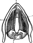
Vocal Cords Seen from above During Quiet Breathing

This illustration shows the vocal cords, seen from above during quiet breathing (A. Thyroid Cartilage;…

Labels: (1 and 2), the ligatures; 3, arytenoid cartilages; 4, thyroid cartilage

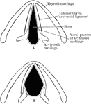
Ligaments of the Vocal Cords

Labels: (1 and 2), the ligatures; 3, arytenoid cartilages; 4, thyroid cartilage

Three laryngoscopic view of the superior aperture of the larynx and surrounding parts. Labels: A, the glottis during the emission of a high note in singing; B, in easy and quite inhalation of air; C, in the state of widest possible dilation, as in inhaling a very deep breath. The diagrams A', B', and C', show in horizontal sections of the glottis in position of the vocal ligaments and arytenoid cartilages in the three several states represented in the other figures. In all the figures, so far as marked, the letters indicate the parts as follows, viz. : l, the base of the tongue; e, the upper free part of the epiglottis; e', the tubercle or cushion of the epiglottis; p h, part of the anterior all of the pharynx behind the larynx; in the margin of the aryteno-epiglottisdean fold w, the swelling of the membrane caused by the cartilages of Wrisberg; s. that of the cartilages of Santorini: a, the tip or summit of the arytenoid cartilages; c v, the true vocal cords r lips of the rima glottidis; c v s, the superior or false vocal cords; between them the ventricle of the larynx; in C, tr, is placed on the anterior wall of the receding trachea, and b indicated the commencement of the two bronchi beyond the bifurcation which may be brought into view in this state of extreme dilation.

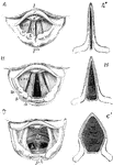
Movement of the Vocal Cords

Three laryngoscopic view of the superior aperture of the larynx and surrounding parts. Labels: A, the…