Clipart tagged: ‘Bladder’
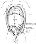
Abdomen of Fetus
The abdominal and thoracic viscera of a five months fetus. The large liver and large size if its left…
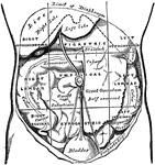
Regions of the Abdomen and their Contents
Regions of the abdomen and their contents (edge of costal cartilages in dotted outline). "For convenience…

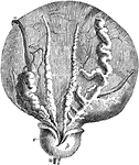
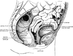
Bladder
View looking into the pelvis from above and somewhat behind. The bladder has been artificially distended.
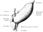
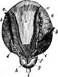
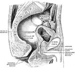
Bladder and Prostate Gland
Dissection of the base of the bladder and prostate gland, showing the vesiculae seminales and vasa deferentia.…
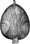
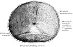
Fibers of the Bladder
The figure on the left shows fibers of the external longitudinal layer. The middle figure shows fibers…
The Bladder
The bladder, a reservoir for urine, is a musculo-membranous sac, situated n the anterior portion of…
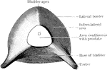
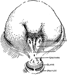
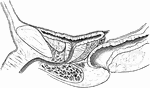
Under Aspect of Male Bladder
The under aspect of the empty male bladder from a subject in which the viscera has been hardened in…
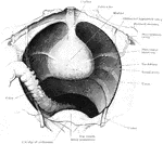
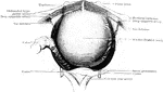
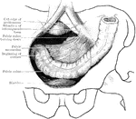
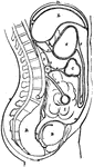
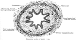
View of Male Pelvis Showing Bladder
View looking into the male pelvis from above and somewhat behind. From a specimen in which the bladder…
Iliac and Pelvic Colons
The iliac and pelvic colons, from a formalin-hardened male body, aged 30. The pelvic colon was usually…
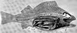
Dissected Fish
"Dissected fish. a, air bladder; b, urinary bladder; b, urinary bladder; br, brain; c, spinal cord;…

Dissected Frog
"Frog with the left side cut away and some of the organs pulled downward. a, aorta leading from the…
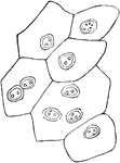
Epithelium of the Bladder
Epithelium of the bladder. Labels: a, one of the cells of the first row; b, a cell of the second row;…
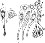
Deep Layer of Bladder Epithelium
Deep layers of the epithelium of the bladder, showing large club-shaped cells above, and smaller, more…
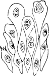
Superficial Layer of Bladder Epithelium
Superficial layer of the epithelium of the bladder. Composed of polyhedral cells of various sizes, each…
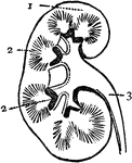
Kidney
Section of Kidney. 1: Body of Kidney; 2: Internal vessels; 3: Ureter, leading to the bladder.
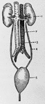
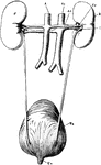
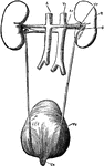
Kidneys
Kidneys and their vessels. 1: Left kidney; 2: Ascending vein; 3: Aorta; 4: Left ureter; 5: Bladder.
The Peritoneum
Diagram of the peritoneum, a serous membrane covering all the contents of the abdominal cavity. Labels:…
Renal Organs
"The renal organs, viewed from behind. R, right kidney; A, aorta; Ar, right renal artery; Vc, inferior…
The Renal Organs Viewed from Behind
The renal organs viewed from behind. labels: R, right kidney; A, aorta; Ar, right erenal artery; Vc,…
Transverse Section of Ureter
In human anatomy, the ureters are muscular tubes that propel urine from the kidneys to the urinary bladder.
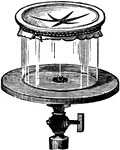
Vacuum
"Over the upper end of a cylindrical receiver, tie tightly a wet bladder or sheet of writing paper and…