Clipart tagged: ‘diaphragm’
Longitudinal Section of Body
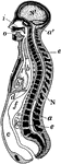
Diagrammatic longitudinal section of the body. Labels: a, the neural tube, with its upper enlargement…
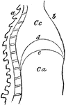
Lateral section of the chest
"A, a muscle which aids in pushing the food down the esophagus; B, esophagus; C,…
Diaphragm
"This is the name applied in anatomy to designate the transverse muscle which, in man and the mammalia…
Diaphragm
"This is the name applied in anatomy to designate the transverse muscle which, in man and the mammalia…
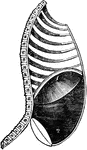
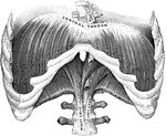
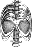
Diaphragm
View of the diaphragm; 1, cavity of the thorax; 2, diaphragm separating the cavity of the thorax from…
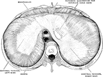
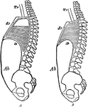
Diaphragm During Expiration
Diaphragm in the state of its greatest ascent in expiration; 2, muscles of the abdomen in action forcing…
Diaphragm During Inspiration
Diaphragm in its state of greatest descent in inspiration; 2, muscles of the abdomen, showing the extend…
The Diaphragm
The diaphragm, which is the principal muscle that act sin breathing. Here you have the cavity of the…
The Diaphragm
Diaphragm of the diaphragm and it's placement in the chest. Let a represent the spinal column,…
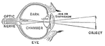
Diagram of the Eye
"Diagram illustrating the Manner in which the Image of an Object is inverted on the Retina." — Blaisedell,…
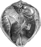
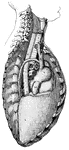
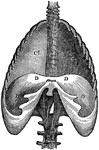
Liver and Diaphragm of a Horse
Posterior view of the liver and diaphragm in situ. Labels: a, left lobe; b, right lobe; c, quadrate…
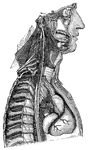
Sympathetic nerve
"The Cervical and Thoracic Portions of the Sympathetic Nerve and their Main Branches. In the center…
The Peritoneum
Diagram of the peritoneum, a serous membrane covering all the contents of the abdominal cavity. Labels:…
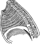
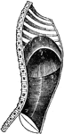
Pleural Sac
Left pleural sac in a subject hardened by formalin injection, opened into by the removal of the costal…
Changes during Respiration
Diagram to show the changes in the sternum, diaphragm, and abdominal wall in respiration. Labels: A,…

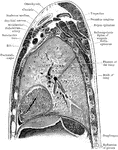
Sagittal Section Through Shoulder and Lung
Sagittal section through left shoulder, lung, and apex of the heart.
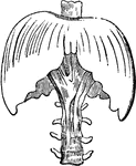
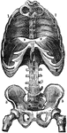
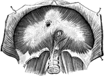
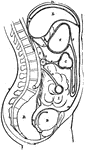
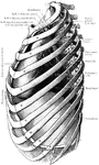
Lower Half of the Thorax
The lower half of the thorax, with four lumbar vertebrae showing the diaphragm from above. Labels: 1,2,3,…
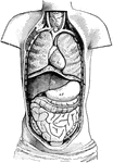
Ventral Cavity of the Body
Diagram of the Body opened from the front to show the contents of the ventral cavity. Labels: d, diaphragm;…