Clipart tagged: ‘human anatomy’
Bones of Arm
Arm of humans; h Humerus or bone of upper arm; r and u Radius and Ulna, or bones of the forearm; c Carpus,…
Arteries and Nerves
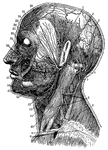
"Superficial arteries and nerves of the face and neck. 1, Temporal artery; 2, artery behind the ear;…
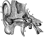
Ear
"Section through right ear. 1, helix; 2, concha; 3, outer passage; 4, 5, 6, semi-circular canals; 7,…
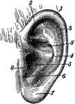
External Ear
"External Ear, or Pinna. 1, helix; 2, fossa of antihelix, or fossa triangularis; 3, fossa of helix,…
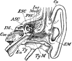
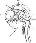
Inner Ear
"Transverse Section through Side Walls of Skull, showing the Inner Parts of the Ear. Co, concha or external…
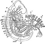
Human Embryo
"Early Human Embryo, giving diagrammatically the principal vessels antecedent to the establishment of…
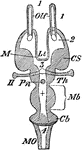
Encephalon
"Diagram of Vertebrate Encephalon ... in longitudinal vertical section. Mb, mid-brain; in front of it…
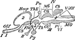
Encephalon
"Diagram of Vertebrate Encephalon ... in horizontal section. Mb, mid-brain; in front of it all is forebrain,…
Eye Cross-Section
"Cross-section of the eye. Parts: co, cornea; I, iris; aq, anterior chamber of aqueous humor; L, lens;…
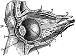
Eye Section
"Section through the left eye, closed. 1, lifting muscle; 2, upper straight muscle; 3, optic nerve;…
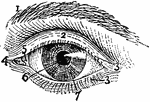
Exterior of Left Human Eye
"Exterior of Left Human Eye. 1, supercilium, or eyebrow; 2, palpebra superior, or upper eyelid; 3, 3,…
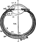
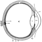
Human Eye
"Diagrammatic horizontal section of the eye of man. c, cornea; ch. choroid (dotted); C. P, ciliary processes;…
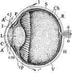
Median Vertical Anteroposterior Section of Eye
"Human Eye, in Median Vertical Anteroposterior Section. (Ciliary processes shown, through not all lying…
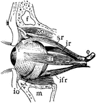
Muscles of Left Eyeball
"Muscles of Left Human Eyeball. so, superior oblique, passing through a trochlea or pulley; io, inferior…
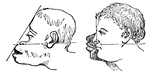
Facial Angles
The facial angle is an angle formed by two imaginary lines; one drawn fom the most prominent part of…
Anterior View of Human Right Femur
"Anterior View of Human Right Femur. ec, external condyle; etu, external tuberosity; ic, internal condyle;…
Youth Femur
"Right Femur of a Youth. E, E, epiphyses; gtr, ltr, greater and lesser trochanter; h, head; et, it,…
Human Fetus
"Diagram of head and brain of human foetus six weeks old (heavy boundaries). The dotted line indicates…
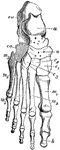
Bones of Human Foot
"Bones of Human Foot, or Pes, the third principal segment of the hind limb, consisting of tarsus, metatarsus,…
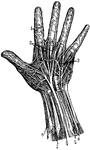
Hand Nerves
"Nerves of the had. 1, Nerves of the skin; 2, tendons; 3, arteries of the palm of the hand; 4, elbow…
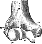
Humerus
"Anterior View, Distal End, of Right Humerus of a Man. H, humerus; epc, epicondyle, or external supracondyloid…
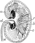
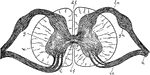
Kidney
Transverse section of the human kidney: "(a) cortex; (b) medulla; (c) small branch of the renal artery;…
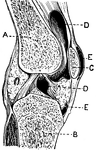
Section of the Knee
This illustration shows a section on the knee (A, Femur; B, Tibia; C, Patella; D, Synovial sac; E, bursæ).…
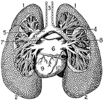
Lungs
"The Lungs. 1, Summit of lungs. 2, Base of lungs. 3, Trachea. 4, Right bronchus. 5, Left bronchus. 6,…

Arm Muscle
A,b,c, deltoid muscle; d, coracobrachialis muscle; r,r, triceps;e,i, extensors of the hand; km, flexor…
Nerve Ganglia (Spinal)
Nerve Ganglia, or Knots (sing. Ganglion; Knot) occur as collections of nerve cells on the course of…
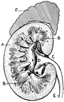
Section of Human Kidney
This illustration shows a section of a human kidney (A, Cortical substance; B, Pyramids; C, Hilum; D,…
Spinal Cord
"Diagram of a cross-section of the spinal cord through the roots of spinal nerves. c, central canal;…

Thorax and Abdomen
"Thorax and abdomen. 1, 1, 1, 1. Muscles of the chest. 2, 2, 2, 2. Ribs. 3, 3, 3. Upper, middle and…