
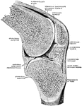
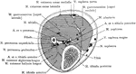
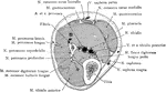
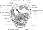
Sagittal Section Through Knee Joint
Right knee joint. Sagittal section through the external condyle of the femur. Mesal half of section,…
Bones of the Knee
Bones of the knee also showing muscle. Labels: s, insertion of the sartorius; g, insertion of the gracilis.
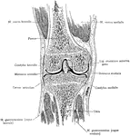
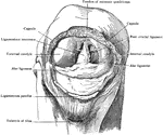
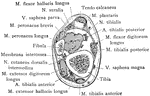
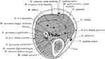
Patella Removed from Knee
Patella removed from right knee, which is strongly flexed to show alar ligaments and ligamentum mucosum.…
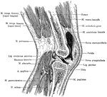
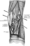
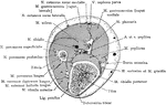
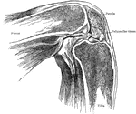
Sagittal Section Through Knee
Sagittal section of the right knee, viewed from the outer side. The joint cavity proper lies to each…
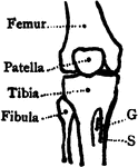
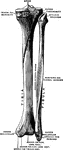
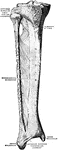
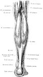
Leg Bones
"Bones of the leg. a, femur; b, tibia; c, fibula; d, tarsal bones; e, metatarsal bones; f, phalanges;…
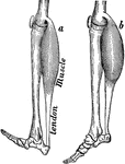
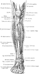
Leg Muscles
The muscles of the front of the leg. 1, tendon of quadriceps; 2, spine tibia; 3, tibialis anticus; 4,…
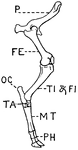
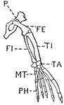
Leg of Ox
This illustration shows the leg of an Ox. P. Pelvis, FE. Femur, TI. Tibia, FI. Fibula, TA. Tarsus, MT.…
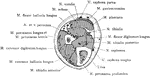
Cross Section of Leg One Inch Above External Malleolus
Section one inch above the external malleolus.
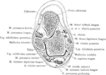
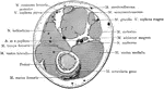
Cross Section Through Leg Three Inches Below Knee Joint
Section through the leg three inches below the right knee joint.
Cross Section of Leg, Two and a Half Inches above Ankle
Section two and a half inches above right ankle joint.
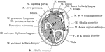
Cross Section Through Leg Two Inches Below Knee Joint
Section through the leg two inches below the right knee joint.
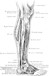
Anterior View of the Superficial Muscles of the Leg
Superficial muscles of the right leg, anterior view.
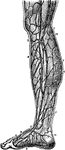
Arteries of the Leg
Arteries of the leg. Labels: 1, extensor propius pollicis; 2, articular arteries; 3, anterior tibia;…
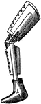
Artificial Leg
In the medical field this is used as a prosthesis or prosthetic limb. This device replaces a missing…

Grooved Artificial Leg
This image depicts an artificial leg having a curved and grooved periphery and supported by the upper…
Lateral View of the Superficial Muscles of the Leg
Superficial muscles of the right leg, lateral view.
Muscles of the leg
"Muscles of the leg showing how they pass into tendons at the ankle." —Davison, 1910
Posterior View of the Superficial Muscles of the Leg
Superficial muscles of the right leg, posterior view.
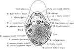
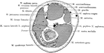
Transverse Section Through the Lower Leg
Transverse section through the lower third of the leg. Labels: a, tibialis anticus; b, extensor longus…
Veins of the Leg
Veins of the leg. Labels: 1, saphenous; 2, collateral branch; 3, anastomosis; 4, internal saphenous;…
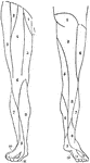
Nerve Supply of Legs
Cutaneous nerve supply of legs. Anterior aspect. 1, ilioinguinal; 2, genitocrural; 3, external cutaneous;…
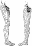
Superficial Lymphatics and Vessels and Nodes of the Legs
Superficial lymphatic vessels and nodes of the right lowers extremity and groin.
Limulus Polyphemus
"Third leg of Limulus polyphemus, showing the division of the fourth segment of the leg by a groove…
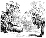
Man with Injured Leg
An illustration of a man sitting in a chair with his leg bandaged and propped up on a chair.
Horse Leg Muscles
Muscles of the anterior limb-external view. Labels: a, antea-spinatus; b, postea-spinatus; c, teres…
Horse Leg Muscles
Muscles of the anterior limb-internal view. Labels: a, subscapularis; b, teres internus; c, coracohumeralis;…
Horse Leg Muscles
External view of the muscles of the anterior limb-showing the deeper ones of the upper region. Labels:…
Horse Leg Muscles
Internal view of the deep muscles of the anterior limb. Labels: a, caput parvum of triceps extensor…
Mystacina Tuberculata
"Thumb and leg and foot of Mystacina tuberculata." —The Encyclopedia Britannica, 1903
Great Nerve
"A Great Nerve (Posterior Tibial) on the Back of the Leg, with its Accompanying Artery of the Same Name."…
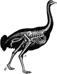
Skeleton of Ostrich
"Shows the powerful legs, small feet, and rudimentary wings of the bird; the obliquity at which the…
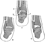
Position of Patella in Relation to Condyles of Femur with Knee Flexed
Showing position of patella in relation to condyles of femur with knee flexed at a right angle.
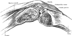
Position of Patella in Relation to Condyles of Femur with Knee Partially Flexed
Showing position of patella in relation to condyles of femur with knee partially flexed.
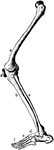
Leg of Seal
This illustration shows the leg of a seal. P. Pelvis, FE. Femur, TI. Tibia, FI. Fibula, TA. Tarsus,…
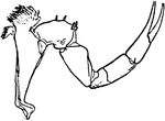
Splint
"When an arm or a leg is broken, it should be kept stretched out straight so that the sharp, broken…
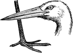
Stork Head and Leg
The head and leg of the stork, a bird in the Ciconiidae family of storks, herons, and egrets.
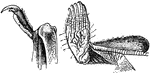
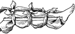
Tendons and Ligaments of Ox Leg
Tendons and ligaments of the left anterior extremity of ox, viewed from external side. Labels: a, flexor…
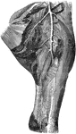
Cross Section Through Thigh Five Inches Above Knee Joint
Section through the lower third of the thigh, five inches above knee joint.
Cross Section Through Thigh Four Inches Above Knee Joint
Section through right thigh, four inches above knee joint.
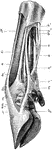
Thigh of a Horse Showing Arteries
Internal view of left thigh-showing the arteries. Labels: 1, femoral; a, profunda femoris; b, superficialis…