The right auricle (atrium) and ventricle of the heart opened, and a part of their right and anterior walls removed, so as to show their interior; 1/2, 1, superior vena cava; 2, inferior vena cava; 2', hepatic veins cut short; 3, right auricle; 3', placed in the fossa ovalis, below which is the Eustachian valve; 3", is placed close to the aperture of the coronary vein; +, +, placed n the auriculo-ventricular groove, where a narrow portion of the adjacent walls of the auricle and ventricle has been preserved; 4, 4, cavity of the right ventricle; the upper figure is immediately below the semilunar valves; 4', large columna carnea or musculus papillaris; 5, 5', 5", tricuspid valve; 6, placed in the interior o the pulmonary artery, a part of the anterior wall of that vessel having been removed, and a narrow portion of it preserved at its commencement where the semilunar valves are attached; 7, concavity of the aortic arch close to the cord of the ductus arteriosus; 8, ascending part of sinus of the arch covered at its commencement by the auricular appendix and pulmonary artery; 9, placed between the innominate and left carotid arteries; 10, appendix of the left auricle; 11, 11, the outside of the left ventricle, the lower figure near the apex.

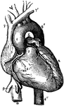
Right Atrium and Ventricle of the Heart

The right auricle (atrium) and ventricle of the heart opened, and a part of their right and anterior…

Front view of the heart and great vessels. The pulmonary artery has been cut short close to its origin. Labels: 1, right ventricle; 2, left ventricle; 3, root of the pulmonary artery; 4, 4', arch of the aorta; 4'', the descending thoracic aorta; 5, part of the right auricle; 6, part of the left auricle; 7, 7', innominate veins joining to form the vena cava superior; 8, inferior vena cava; 9, one of the large hepatic veins; X, placed in the right auriculo-ventricular groove, points to the right or posterior coronary artery; X, X, placed in the anterior interventricular groove, indicate the left or anterior coronary artery.

Heart

Front view of the heart and great vessels. The pulmonary artery has been cut short close to its origin.…

A representation of the heart as it really appears showing the front view. At <em>a</em> is the right auricle that receives blood from all pats of the body by two large veins h and i. At <em>b</em> is the right ventricle, which receives the blood from the auricle and sends it to the lungs by the pulmonary artery <em>f</em>. At <em>c</em> is the left auricle, which receives the blood from the lungs by the pulmonary veins <em>g, g, g</em>. At <em>d</em> is the left ventricle which receives blood from the auricle and forces it all over the body through the aorta <em>e</em>.

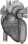
Heart, Front View of

A representation of the heart as it really appears showing the front view. At a is the right…

Diagram of the interior of the heart. Labels: A, aorta; PA, pulmonary artery; VCI and VCS, vena cava inferior and vena cava superior; T, tricuspid, and M, mitral valve. The other letters same as in preceding figure.

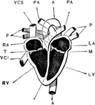
Interior of the Heart

Diagram of the interior of the heart. Labels: A, aorta; PA, pulmonary artery; VCI and VCS, vena cava…

Section of lung, showing small air tubes and branch of pulmonary artery.

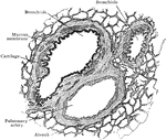
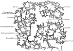
Section of Lung Showing Air Tubes

Section of lung, showing small air tubes and branch of pulmonary artery.

Section of lung, showing general relations of division of air tubes.

Section of Lung Showing General Relations of Divisions of Air Tubes

Section of lung, showing general relations of division of air tubes.