Spine
Diagram on frozen section, showing relations of bodies and spines of vertebrae to levels at which spinal…
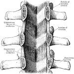
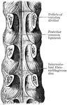
Ligamenta Subflava of the Spine
Ligamenta subflava as seen from the front after removal of he bodies as the vertebrae by sawing through…
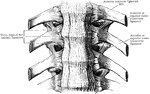
Ligaments of the Spine
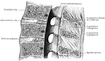
Anterior common ligament of the vertebral column, and the costo vertebral joints as seen from in front.
Spinet
"The spinet is a keyed musical instrument much in use from 1500 to 1760. It derived its name from the…
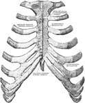
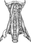
Sternum and Ribs
Sternum and ribs with ligaments, from in front. In the right half of the figure the most anterior layer…
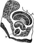
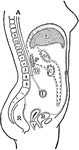
Vertical Median Section of the Trunk
Diagram of vertical median section of abdomen. The fine dots represent the great sac of the peritoneum,…
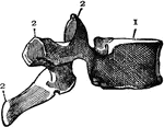
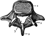
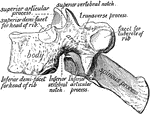
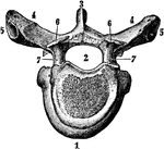
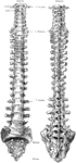
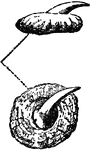
Two Views of a Vertebra
Two views of vertebra. 24 vertebrae make up the spinal column. On the left figure, a is the…
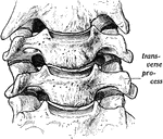
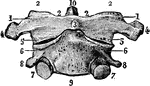
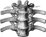
Upper Two Vertebrae
The upper two vertebrae of the spinal column, involved in head movement. In performing the rotary motion…
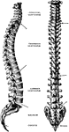
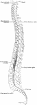
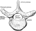
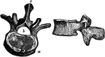
Lateral and Dorsal View of the Vertebral Column
The spinal column, right lateral view and dorsal view.
Great Weaver
"It is about twelve inches long, lives in deep water, and is noted for inflicting serious wounds with…