Pterylosis of Cypselus Apus
| View Cart ⇗ | Info

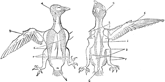
“Fig. 24. - Pterylosis of Cyoselus apus, drawn by Coues after Nitzsch; right hand upper, left hand lower, surface. 1 spinal tract; 2. humeral; 3. femoral; 4. capital; 5. alar; 6. caudal; 7. crural; 8. ventral.” Elliot Coues, 1884
Keywords
birds, capital, ornithology, bird anatomy, feathers, ventral, femoral, Crural, Caudal, humeral, pterylosis, feather structures, Cyoselus apus, spinal tract, alar, external bird partsGalleries
Bird AnatomySource
Elliot Coues Key to North American Birds (Boston, MA: Estes and Lauriat, 1884)
Downloads
2400×1197, 323.3 KiB
1024×510, 57.5 KiB
640×319, 31.1 KiB
320×159, 10.8 KiB