This human anatomy ClipArt gallery offers 148 illustrations of general dissected or cross-sectional views of the human body that do not focus on a particular system.

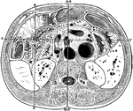
Thorax and Abdomen
"Thorax and abdomen. 1, 1, 1, 1. Muscles of the chest. 2, 2, 2, 2. Ribs. 3, 3, 3. Upper, middle and…
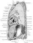
Side View of the Thorax and Part of the Abdomen
Lateral, sagittal section through the left thorax and upper portion of abdomen, viewed from the left.…
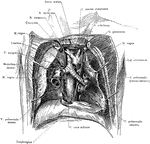
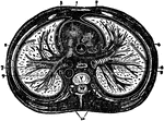
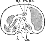
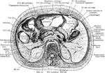
Dissection of the Thorax
Topography of the retrocardiac structures of the mediastinum, after the removal of the heart and pericardium.
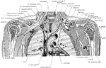
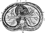
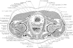
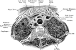
Frontal Section Through the Thorax
Frontal section through the thorax, passing through the mediastinum and the middle of the humeral heads.
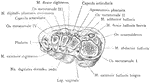
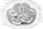
The Thorax
Diagram of a transverse section of the thorax. Labels: 1, anterior mediastinum; 2, internal mammary…
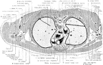
Transverse Section of the Thorax
Transverse section of the thorax. Labels: 1, anterior mediastinum; 2, internal mammary vessels; 3, triangularis…
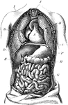
Torso
The human torso. Labels: A, the heart; B, the lungs drawn aside to show the internal organs; C, the…
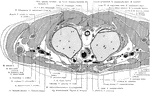
Cross Section of the Trunk above Pubic Symphysis
Section through the pelvis immediately above the pubic symphysis.
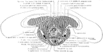
Cross Section of the Trunk above the Manubrium Sterni
Section immediately above the manubrium sterni.
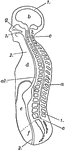
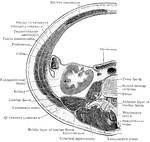
Trunk and Head of Human Body
Diagrammatic longitudinal section of the trunk and head. Labels: 1,1, the dorsal cavity; a, the spinal…
Cross Section Through the Trunk at the Neck
Section through the neck immediately above the shoulders.
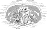
Cross Section of the Trunk Below Umbilicus
Section through the abdomen immediately below the umbilicus and the superior iliac crest.
Horizontal Section Through Trunk
Diagram of horizontal section through upper part of 1st lumbar vertebra. The fine dots represent the…
Horizontal Section Through the Trunk
Horizontal section through the trunk at the level of the first lumbar vertebra showing descriptive terms.…
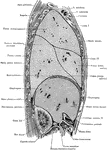
Side View of the Trunk
Sagittal section through the trunk, 6 cm to the right of the median plane, viewed from the right. Note…
Transverse Section Through
Transverse section through the abdomen, opposite the second lumbar vertebra.
Vertical Median Section of the Trunk
Diagram of vertical median section of abdomen. The fine dots represent the great sac of the peritoneum,…
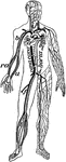
Veins and Arteries of the Body
Chief veins and arteries of the body. Labels: a, place of the heart; the veins are in the back. On the…
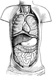
Ventral Cavity of the Body
Diagram of the Body opened from the front to show the contents of the ventral cavity. Labels: d, diaphragm;…
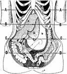
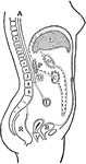
Position of the Viscera in the Condition of Visceroptosis
Showing the position of the viscera in the condition of visceroptosis (Glenard's disease). Labels: A,…
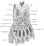
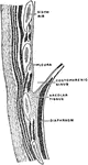
Transverse Section Through the Wrist
Transverse section through the wrist, showing the annular ligaments and the canals for the passage of…