This human anatomy ClipArt gallery offers 148 illustrations of general dissected or cross-sectional views of the human body that do not focus on a particular system.
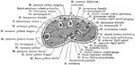
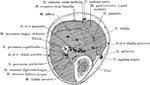
Cross Section on a Line with the Palmar Arch of the Hand
Section on a line with the deep palmar arch of the right hand.

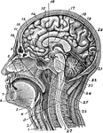
Sagittal Section of the Head and Neck
Sagittal median section of the head and neck. The head is thrown backward into complete extension which…
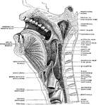
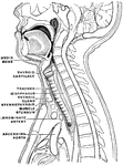
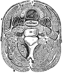
Section of the Head and Neck
Section of head and neck from front to back. Labels: 1, windpipe; 2, larynx; 3, spinal marrow; 4, pharynx;…

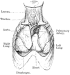
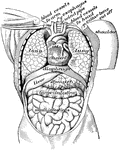
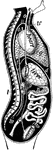
Heart and Lungs
View of heart and lungs in situ. The front portion of the chest wall, and the outer or parietal layers…
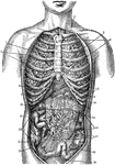
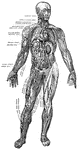
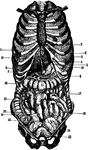
Internal Anatomy
1. Collar bone 2. Left Lung 3. Breast Bone 4. Right Lung 5. Ribs 6. Right lobe of the liver 7. Left…
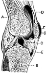
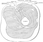
Section of the Knee
This illustration shows a section on the knee (A, Femur; B, Tibia; C, Patella; D, Synovial sac; E, bursæ).…
Cross Section of Leg One Inch Above External Malleolus
Section one inch above the external malleolus.
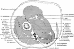
Cross Section Through Leg Three Inches Below Knee Joint
Section through the leg three inches below the right knee joint.
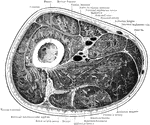
Cross Section Through Leg Two Inches Below Knee Joint
Section through the leg two inches below the right knee joint.
Front of Leg
Front of leg. Labels: 1, external popliteal nerve; 2, anterior tibial artery; 3, musculocutaneous nerve;…

Grooved Artificial Leg
This image depicts an artificial leg having a curved and grooved periphery and supported by the upper…
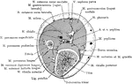
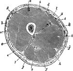
Transverse Section of the Leg
Transverse section of the thigh below the lesser trochanter. The femoral artery, vein, and nerve are…
Transverse Section Through Leg
Transverse section at the middle of the leg. In front of the interosseous membrane are the anterior…
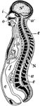
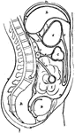
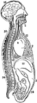
Longitudinal Section of the Body
Diagrammatic longitudinal section of the Body. Labels: a, the neural tube, with its upper enlargement…
Muscles and Blood Vessels
"Principal Muscles on the Right, Certain Organs of the Chest and Abdomen, and the Larger Blood Vessels…
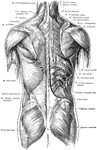
Posterior View of the Muscles of the Trunk
Superficial and deep muscles of the trunk. The latissimus dorsi and trapezius on the right side have…
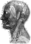
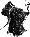
Neck
1. Temporal Artery 2. Artery behind the ear 3. Occipital Artery 4. Greater occipital nerve 5. Smaller…

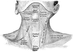

Section of the Neck
Section of the neck at about the level of the sixth cervical vertebra. Showing the arrangement of the…
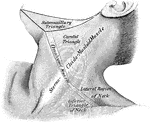
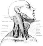
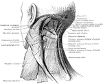
The Side of the Neck
The side of the neck, showing the nerves and blood vessels. Labels: 1, occipital artery; 2, facial vein;…
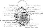
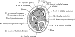
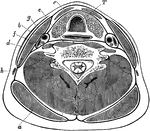
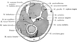
The Transverse Section of the Neck
A transverse section of the neck. The separate muscles as they are arranged in layers, with their investing…
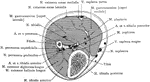
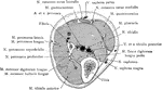
Transverse Section of the Neck
Transverse section through the lower part of the neck, to show the arrangement of the cervical fascia.…
Nerve Ganglia (Spinal)
Nerve Ganglia, or Knots (sing. Ganglion; Knot) occur as collections of nerve cells on the course of…
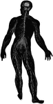
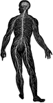
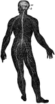
Diagram of the Human Nervous System
Diagram illustrating the general arrangement of the cerebrospinal nervous system.
View of Organs from the Side
The chief organs of the body from the side. Labels: a, arch of the aorta or main artery of the trunk;…
The Peritoneum
The peritoneum is a large serous membrane, which forms in the male a closed sac, the parietal layer…
The Peritoneum
Diagram of the peritoneum, a serous membrane covering all the contents of the abdominal cavity. Labels:…

Sagittal Section Through Shoulder and Lung
Sagittal section through left shoulder, lung, and apex of the heart.
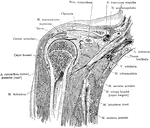
Frontal Section Through the Shoulder
Frontal section of the right shoulder through the middle of the humeral head. Section passes through…
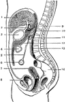
Side View of the Body
A side view of the two great cavities of the body and their organs. 1: The mouth. 2: The thorax. 3:…
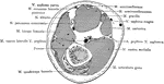
Cross Section Through Thigh Five Inches Above Knee Joint
Section through the lower third of the thigh, five inches above knee joint.
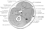
Cross Section Through Thigh Four Inches Above Knee Joint
Section through right thigh, four inches above knee joint.


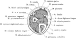
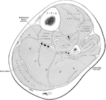
Transverse Section Through the Thigh
Transverse section through the middle of the thigh. Labels: a, Rectus femoris; b, vastus externus; c,…
Section Across the Forearm
Diagram showing the position of the thoracic and abdominal organs. labels: 1, lower border of right…
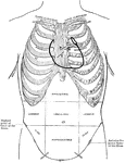
Diagram of Thoracic and Abdominal Regions
A diagram of the thoracic and abdominal regions. Labels: A, aortic valve; M, mitral valve; p, pulmonary…
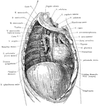
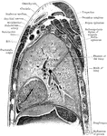
Thoracic Cavity after Removal of Lung
Deep structures of the right thoracic cavity, after removal of the right lung.
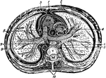
Thorax
"The transverse section of the thorax. 1, anterior mediastinum; 2, internal mammary vessels; 3, triangularis…