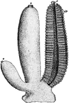
The Sponges ClipArt gallery includes 48 illustrations of sponges. Sponges, also called poriferans, are in the phylum porifera and are all sessile animals that live and feed attached to the bottom of the sea.

"Simple sponge (Ascetta primordialis). Note the vase-like form, the apical osculum, the inhalant pores in the walls." -Thomson, 1916

A. Primordialis

"Simple sponge (Ascetta primordialis). Note the vase-like form, the apical osculum, the inhalant pores…

Diagram showing the Ascon type of sponge. ec., ectoderm; r.c., radiating canals; i.p., internal pores; mes., mesenchyma, e.p., external pores.

Ascon Sponge

Diagram showing the Ascon type of sponge. ec., ectoderm; r.c., radiating canals; i.p., internal pores;…

"Diagram showing types of canal system. --After Korschelt and Heider. The flagellate regions are dark throughout, the mesogloea is dotted, the arrows show the direction of the currents. All the figures represent cross-sections through the wall. A. Simple Ascon type (Ec., outer layer; En., inner layer; Mg., mesogloea). B. Sycon type, with flagellage radial chambers (r.c.). CC. Leucon type, with flagellate side aisles on the main radial chambers. D. Still more complex type, with small flagellate chambers (f.ch.)." -Thomson, 1916

Sponge Canal System

"Diagram showing types of canal system. --After Korschelt and Heider. The flagellate regions are dark…
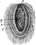
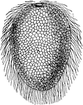
"Gastrula of a Chalk-sponge (Olynthus). A, external view." -Whitney, 1911

Chalk Sponge Gastrula

"Gastrula of a Chalk-sponge (Olynthus). A, external view." -Whitney, 1911

"Gastrula of a Chalk-sponge (Olynthus). B, longitudinal section through the axis: g, primitive intestine; o, blastopore; i, inner cell-layer of the body-wall; e, outer cell-layer." -Whitney, 1911

Chalk Sponge Gastrula

"Gastrula of a Chalk-sponge (Olynthus). B, longitudinal section through the axis: g, primitive intestine;…

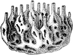
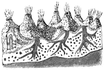
A sponge colony.

Sponge Colony

A sponge colony.

"Connective tissue of sponge." — Encyclopedia BSponge Diver Counting Cardsritannica, 1893

Connective Tissue

"Connective tissue of sponge." — Encyclopedia BSponge Diver Counting Cardsritannica, 1893

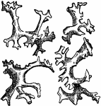
Isolated spicular bodies of an extinct silicious sponge. (<em>Epistomella clivosa</em>).

Epistomella Clivosa

Isolated spicular bodies of an extinct silicious sponge. (Epistomella clivosa).

A sea sponge in the shape of a human glove.

Neptune's Glove

A sea sponge in the shape of a human glove.

A modern sponge with silicious skeletal structure, and basal glass fibers for fixation (<em>Holtenia carpenteri</em>).

Holtenia Carpenteri

A modern sponge with silicious skeletal structure, and basal glass fibers for fixation (Holtenia

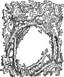
Part of the skeletal structure of a lithisdid silicious sponge.

Jereiea Polystoma

Part of the skeletal structure of a lithisdid silicious sponge.

"Diagrammatic representation of development of Oscarella lobularis. Bl., Free-swimming blastula with flagella; G., gastrula settled down. Next figure shows folding of inner layer (En.); Ec., outer layer. Lowest figure shows radial chambers (R.C.); Mesogloea (Mg.); inhalant pore (P.); exhalant osculum (O.)." -Thomson, 1916

O. Lobularis

"Diagrammatic representation of development of Oscarella lobularis. Bl., Free-swimming blastula with…

"Section of a sponge. Showing inhalant canals, flagellate chambers, a gastrula forming in the mesogloea, etc." -Thomson, 1916

Sponge Section

"Section of a sponge. Showing inhalant canals, flagellate chambers, a gastrula forming in the mesogloea,…
Part of the outer wall of this Calcolynthus is cut away. This is a simple sponge.

simple sponge

Part of the outer wall of this Calcolynthus is cut away. This is a simple sponge.

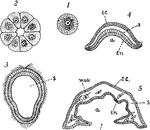
"Diagrams to illustrate the development of one of the simpler types of sponge: I, the egg; 2, section of 16- to 32-celled stage; 3, section of later stage, a ciliated larva (blastula); 4, gastrula; 5, section through older larva which has become attached by the end containing the blastopore. New openings break through by the coalescence and perforation of the ectoderm and entoderm, and and a form results... a, archenteron; bl., blastopore; ec., ectoderm; en., entoderm; mes., mesenchyma; s, segmentation cavity." -Galloway, 1915

Simple Sponge Development

"Diagrams to illustrate the development of one of the simpler types of sponge: I, the egg; 2, section…
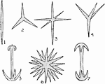
"Sponge spicules. 1, Monaxon; 2, triod; 3, triaxon; 4, tetraxon; 5, anchor; 6, polyaxon; 7, a kind of amphidisc." -Thomson, 1916

Sponge Spicules

"Sponge spicules. 1, Monaxon; 2, triod; 3, triaxon; 4, tetraxon; 5, anchor; 6, polyaxon; 7, a kind of…
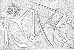
Spicules may be made of silica or calcium carbonate, and vary in shape from simple rods to three-dimensional "stars" with up to six rays. Spicules are produced by sclerocyte cells, and may be separate, connected by joints, or fused.

Sponge Spicules

Spicules may be made of silica or calcium carbonate, and vary in shape from simple rods to three-dimensional…

In this simple sponge, water enters minute holes in the sides and passes out of the opening at the top of the tube.

Sponge

In this simple sponge, water enters minute holes in the sides and passes out of the opening at the top…
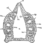
In this simple sponge, part of the outer wall is cut away to show the inside.

Sponge

In this simple sponge, part of the outer wall is cut away to show the inside.

A cross-section of a simple tubular sponge.

Sponge

A cross-section of a simple tubular sponge.

A diagram of a commercial sponge.

Sponge

A diagram of a commercial sponge.

A typical sea sponge growing on the ocean floor

Sponge

A typical sea sponge growing on the ocean floor

An animal of the porifera class, having pores in the body-wall and being without tentacles.

Sponge

An animal of the porifera class, having pores in the body-wall and being without tentacles.

"Diagram of simple type of sponge. c, cloaca; ch, chambers, lined with flagellate entoderm; e.p., external pores; i.p., internal pores; mes., mesenchyma; o, osculum; r.c., radiating canals. In the adult sponge the canals and flagellate chambers become much more complex than figured here." &mdash; Galloway

Sponge

"Diagram of simple type of sponge. c, cloaca; ch, chambers, lined with flagellate entoderm; e.p., external…

The sponge, a many celled animal, begins its life as a single-cell, the egg.

Sponge

The sponge, a many celled animal, begins its life as a single-cell, the egg.

The young sponge, a many celled animal, that begins its life as a single-cell, the egg.

Sponge

The young sponge, a many celled animal, that begins its life as a single-cell, the egg.

The young sponge, a many celled animal, that begins its life as a single-cell, the egg. A section of young larva.

Sponge

The young sponge, a many celled animal, that begins its life as a single-cell, the egg. A section of…

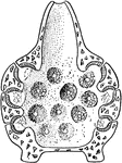
Calcolynthus primigenius, a simple sponge with the wall removed to show the inside. It is hollow, attached at the base with an opening at the top. There are many small holes though the wall with water constantly entering and passing out through the mouth at the top.

Sponge

Calcolynthus primigenius, a simple sponge with the wall removed to show the inside. It is hollow, attached…
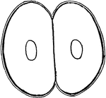
The sponge, a many celled animal, begins its life as a single-cell, the egg. First division in into two cell stage.

Sponge Develoment

The sponge, a many celled animal, begins its life as a single-cell, the egg. First division in into…
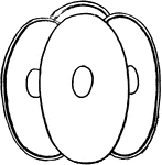
The sponge, a many celled animal, begins its life as a single-cell, the egg. Second division into the four cell stage.

Sponge Develoment

The sponge, a many celled animal, begins its life as a single-cell, the egg. Second division into the…
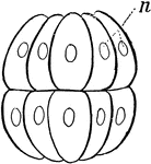
The sponge, a many celled animal, begins its life as a single-cell, the egg. Fourth division into a sixteen cell stage. This repeated process give the young sponge the general form of Pandorina.

Sponge Develoment

The sponge, a many celled animal, begins its life as a single-cell, the egg. Fourth division into a…
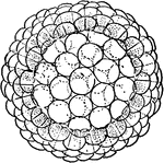
The sponge, a many celled animal, begins its life as a single-cell, the egg. The continued division of its cells result in the formation of a hollow globe of cells called the blastula, similar to Volvox, and at this point the young larva leaves the parent.

Sponge Develoment

The sponge, a many celled animal, begins its life as a single-cell, the egg. The continued division…
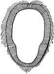
The sponge, a many celled animal, that begins its life as a single-cell, the egg. The continued division of its cells result in the formation of a hollow globe of cells called the blastula, similar to Volvox, and at this point the young larva leaves the parent.

Sponge Develoment

The sponge, a many celled animal, that begins its life as a single-cell, the egg. The continued division…

The sponge, a many celled animal, begins its life as a single-cell, the egg. After division of cells and formation of blastula, the young larva leaves the parent. The next transformation consists of a pushing in of one side. This depression deepens and gradually results in the formation of a two layered sac know as the gastrula.

Sponge Develoment

The sponge, a many celled animal, begins its life as a single-cell, the egg. After division of cells…

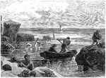
An illustration of men collecting sponges.

Sponge Fishing

An illustration of men collecting sponges.

An illustration of a sponge fossil.

Sponge Fossil

An illustration of a sponge fossil.

A Cretaceous sponge with silicious skeleton retaining its form. (<em>Ventriculites simplex</em>).

A Cretaceous Sponge

A Cretaceous sponge with silicious skeleton retaining its form. (Ventriculites simplex).

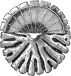
A beautiful Glass Sponge called the "Venus Flower Basket". It grows in the deep sea near the Philippine Islands.

Glass Sponge

A beautiful Glass Sponge called the "Venus Flower Basket". It grows in the deep sea near the Philippine…
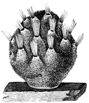
"A glace at a piece of common sponge will show that its surface is everywhere perforated with an infinite number of minute holes, among which a considerable number of large openings are scattered. When a sponge is examined in a living state, a rapid stream of water may be observed issuing constantly from these larger orifices." &mdash; Goodrich, 1859

Living sponge, magnified

"A glace at a piece of common sponge will show that its surface is everywhere perforated with an infinite…
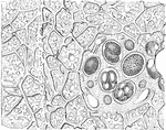
An illustration of the lower portion of a sponge.  O, OS, and M are illustrations of sponge eggs magnified forty times.  The sponges or poriferans (from Latin porus "pore" and ferre "to bear") are animals of the phylum Porifera. Their bodies consist of an outer thin layer of cells, the pinacoderm and an inner mass of cells and skeletal elements, the choanoderm. Sponges do not have nervous, digestive or circulatory systems. Instead most rely on maintaining a constant water flow through their bodies to obtain food and oxygen and to remove wastes, and the shapes of their bodies are adapted to maximize the efficiency of the water flow.

Lower Section of Sponge

An illustration of the lower portion of a sponge. O, OS, and M are illustrations of sponge eggs magnified…

In zoology a skeleton is any fairly rigid structure of an animal, irrespective of whether it has joints and irrespective of whether it is biomineralized. The mesohyl functions as an endoskeleton in most sponges, and is the only skeleton in soft sponges that encrust hard surfaces such as rocks. More commonly the mesohyl is stiffened by mineral spicules, by spongin fibers or both. Portion of a wall of sponge (fig 17) showing the outer layer (e), the inner layer (i), consisting of collared cells, and the middle layer (m), consisting of irregular cells.

Skeleton of Sponge

In zoology a skeleton is any fairly rigid structure of an animal, irrespective of whether it has joints…
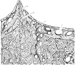
An illustration of the upper portion of a sponge: p, Pore; s, Subdermal cavity; c1, chief fiver of the skeleton; c2, connecting film. The sponges or poriferans (from Latin porus "pore" and ferre "to bear") are animals of the phylum Porifera. Their bodies consist of an outer thin layer of cells, the pinacoderm and an inner mass of cells and skeletal elements, the choanoderm. Sponges do not have nervous, digestive or circulatory systems. Instead most rely on maintaining a constant water flow through their bodies to obtain food and oxygen and to remove wastes, and the shapes of their bodies are adapted to maximize the efficiency of the water flow.

Upper Section of Sponge

An illustration of the upper portion of a sponge: p, Pore; s, Subdermal cavity; c1, chief fiver of the…

Sea sponges in action

Sponges

Sea sponges in action

"Vertical section of a fresh-water sponge (Spongilla), showing the arrangement of the canal-system. C, ciliated chambers; DP, dermal pores; Ex, excurrent canals; GO, openings of the excurrent canals; PG, paragastric cavity; SD, subdermal cavities; O, osculum." -Parker, 1900

Spongilla

"Vertical section of a fresh-water sponge (Spongilla), showing the arrangement of the canal-system.…

Sycon ciliatum, a sponge.

Sycon Ciliatum

Sycon ciliatum, a sponge.

Sycon gelatinosum, a sponge.

Sycon Gelatinosum

Sycon gelatinosum, a sponge.

"Transverse section through the wall of a cylinder (parallel with the course of the canals), showing one incurrent canal (IC), and one radial (R) throughout their length; sp, triradiate spicules; sp', oxeote spicules of dermal cortex (dc); sp'', tetraradiate spicules of gastral cortex (gc); ec, ectoderm; en, endoderm; pm, pore membrane; pp, prosopyles; ap, apopyle; di, diaphragm; exc, excurrent passage; PG, paragastric cavity; em, early embryo; em, late embryo. The arrows indicate the course of the water through the sponge." -Parker, 1900

Sycon Gelatinosum

"Transverse section through the wall of a cylinder (parallel with the course of the canals), showing…
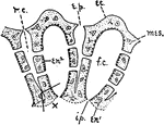
Diagram showing the Sycon type of sponge. ec., ectoderm; r.c., radiating canals; i.p., internal pores; mes., mesenchyma, e.p., external pores; en1, entoderm; en2, flagellate entoderm; f.c., flagellate chambers.

Sycon Sponge

Diagram showing the Sycon type of sponge. ec., ectoderm; r.c., radiating canals; i.p., internal pores;…