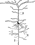
A neuron from the cerebral cortex. The axis-cylinder process, dendrites and collaterals are marked 1, 2, and 3, respectively.

Neuron from the Cerebral Cortex

A neuron from the cerebral cortex. The axis-cylinder process, dendrites and collaterals are marked 1,…
Semidiagrammatic view of the cerebrospinal axis.

Cerebrospinal Axis

Semidiagrammatic view of the cerebrospinal axis.

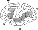
Lateral view of a brain hemisphere; cortical area V, damage to which produces "mind blindness" (word blindness); cortical area H, damage to which produced "mind deafness" (word deafness); cortical area S, damage to which caused the loss of audible speech; cortical area W, damage to which abolished the power of writing.

Association Area of the Brain

Lateral view of a brain hemisphere; cortical area V, damage to which produces "mind blindness" (word…
Front view of the heart.

Front View of the Heart

Front view of the heart.

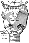
Laryngeal cartilages and ligaments from in front.

Cartilage of the Larynx

Laryngeal cartilages and ligaments from in front.

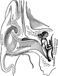
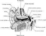
Vertical section through the external auditory meatus and tympanum, passing in front of the fenestra ovalis.

Ear Showing External Auditory Meatus

Vertical section through the external auditory meatus and tympanum, passing in front of the fenestra…
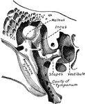
Chains of small bones and their ligaments, seen from the front in a vertical, transverse section of the tympanum.

Small Bones and Ligaments of the Ear

Chains of small bones and their ligaments, seen from the front in a vertical, transverse section of…

Lateral and posterior views of the vertebral column.

Lateral and Posterior View of the Vertebral Column

Lateral and posterior views of the vertebral column.
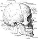
Skull and mandible, lateral view.

Skull and Mandible

Skull and mandible, lateral view.

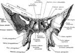
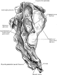
Sphenoid bone, viewed from in front.

Sphenoid Bone

Sphenoid bone, viewed from in front.

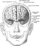
The brain in relation to the skull and face, anterior view.

Brain in Relation to Skull and Face

The brain in relation to the skull and face, anterior view.

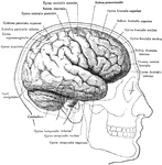
The brain in relation to the skull and face, lateral view.

Brain in Relation to Skull and Face

The brain in relation to the skull and face, lateral view.

Vertical section through the right external acoustic canal, viewed from in front.

Auditory Canal of the Ear

Vertical section through the right external acoustic canal, viewed from in front.
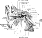
General view of the right external ear and middle. The external ear has been opened by a vertical section carried obliquely lateralward and dorsalward.

External Ear and Middle Ear

General view of the right external ear and middle. The external ear has been opened by a vertical section…
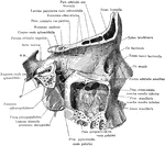
Right pterygopalatine fossa, from without. The greater portion of the ala magna oss. sphenoid., of the lateral part of the corpus maxillae and of the pars orbit. oss. front. have been removed by a sagittal section, exposed the medial wall of the sinus maxillaris and of the orbit. Labels: **, indicate a probe through the Foramen rotundum; *, indicates a probe through the Canalis pterygoideus.

Pterygoid Fossa and Maxillary Sinus

Right pterygopalatine fossa, from without. The greater portion of the ala magna oss. sphenoid., of the…

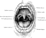
The oral cavity seen from in front.

Oral Cavity

The oral cavity seen from in front.

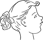
Profile view of a young girl.

Profile of a young girl

Profile view of a young girl.

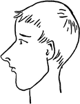
Profile view of a young boy.

Profile of a young boy

Profile view of a young boy.

The Cuban pine, which occurs mostly in the West Indies and South America, as well as the Gulf States of America.

Cuban pine (pinus cubensis Griseb.). Two-thirds natural size. cone scales, dorsal view

The Cuban pine, which occurs mostly in the West Indies and South America, as well as the Gulf States…

The Cuban pine, which occurs mostly in the West Indies and South America, as well as the Gulf States of America.

Cuban pine (pinus cubensis Griseb.). Two-thirds natural size. cone scales, ventral view

The Cuban pine, which occurs mostly in the West Indies and South America, as well as the Gulf States…

The Cuban pine, which occurs mostly in the West Indies and South America, as well as the Gulf States of America.

Cuban pine (pinus cubensis Griseb.). Two-thirds natural size. seed wings dorsal view

The Cuban pine, which occurs mostly in the West Indies and South America, as well as the Gulf States…

The Cuban pine, which occurs mostly in the West Indies and South America, as well as the Gulf States of America.

Cuban pine (pinus cubensis Griseb.). Two-thirds natural size. seed wings ventral view

The Cuban pine, which occurs mostly in the West Indies and South America, as well as the Gulf States…

The shortleaf pine is mostly associated with with deciduous-leaved trees, often the predominant forest growth in parts of North America.

Shortleaf pine (pinus echinata Mill.). Natural size. cone scales, dorsal view

The shortleaf pine is mostly associated with with deciduous-leaved trees, often the predominant forest…

The shortleaf pine is mostly associated with with deciduous-leaved trees, often the predominant forest growth in parts of North America.

Shortleaf pine (pinus echinata Mill.). Natural size. cone scales, ventral view

The shortleaf pine is mostly associated with with deciduous-leaved trees, often the predominant forest…

The loblolly pine, also known as slash-pine is a common pine tree in the Virginias and Carolinas.

Loblolly pine (pinus toeda L.). Two-thirds natural size. detached cone scales dorsal view

The loblolly pine, also known as slash-pine is a common pine tree in the Virginias and Carolinas.

The loblolly pine, also known as slash-pine is a common pine tree in the Virginias and Carolinas.

Loblolly pine (pinus toeda L.). Two-thirds natural size. detached cone scales ventral view

The loblolly pine, also known as slash-pine is a common pine tree in the Virginias and Carolinas.
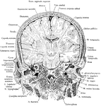
Frontal section of the head, passing through external and internal auditory meatus, as seen from in front.

Frontal Section of Head

Frontal section of the head, passing through external and internal auditory meatus, as seen from in…
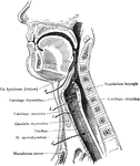
Operative approaches through the front of the neck to the larynx, pharynx, and trachea. a: Approach to pharynx between the hyoid and the thyroid cartilages. b: Approach to the larynx between the thyroid and cricoid cartilages. c: approach to the larynx below the cricoid cartilage and above the isthmus of the thyroid gland. d: Approach for low tracheotomy.

Upper Respiratory Tract

Operative approaches through the front of the neck to the larynx, pharynx, and trachea. a: Approach…
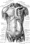
Superficial and deep muscles of the trunk. The sternocleidomastoid, pectoralis major, anterior portion of deltoid, the external oblique, aponeurosis of the internal oblique, tensor fasciae latae, sartorius, rectus femoris, iliopsoas, pectineus, adductor longus and the gracilis muscles of the left side have been cut away to expose the underlying muscles. Anterior View.

Anterior View of the Muscles of the Trunk

Superficial and deep muscles of the trunk. The sternocleidomastoid, pectoralis major, anterior portion…

Superficial and deep muscles of the trunk. The latissimus dorsi and trapezius on the right side have been cut away to expose the underlying muscles. Posterior View.

Posterior View of the Muscles of the Trunk

Superficial and deep muscles of the trunk. The latissimus dorsi and trapezius on the right side have…
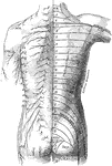
The distribution of cutaneous nerves n the back of the trunk. On the left side the distribution of the several nerves is represented, the letters indicating their nomenclature. ACR.; Posterior supraclavicular branches from cervical plexus; CIRC: Cutaneous branches of axillary nerve; E.C.: Lateral femoral cutaneous nerve, L.1: Lateral cutaneous branch of iliohypogastric nerve; S.1; Lateral branches of sacral nerves; T: T.1-12: Cutaneous branches of the posterior rami of the thoracic nerves. On the right side is a schematic representation of the areas supplied by the above nerves, the numeral and letter s indicate the spinal origins of the nerves distributed to each area.

Posterior View of the Cutaneous Nerves of Trunk

The distribution of cutaneous nerves n the back of the trunk. On the left side the distribution of the…
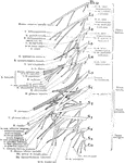
Right lumbar and sacral plexuses, schematic, viewed from in front The darkly shaded trunks are derivatives of the dorsal half of the plexus.

Lumbar and Sacral Nerve Plexuses

Right lumbar and sacral plexuses, schematic, viewed from in front The darkly shaded trunks are derivatives…
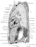
Lateral, sagittal section through the left thorax and upper portion of abdomen, viewed from the left. The plane of the section lies 4 cm to the left of the median plane.

Side View of the Thorax and Part of the Abdomen

Lateral, sagittal section through the left thorax and upper portion of abdomen, viewed from the left.…
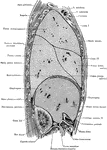
Sagittal section through the trunk, 6 cm to the right of the median plane, viewed from the right. Note the close proximity of the duodenum and colon to the right kidney.

Side View of the Trunk

Sagittal section through the trunk, 6 cm to the right of the median plane, viewed from the right. Note…

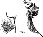
The side view of a human neck.

Neck

The side view of a human neck.

Frontal section through the right kidney and adjacent structures showing the renal fasciae and fatty layers, viewed from in front.

Frontal Section Through Kidney

Frontal section through the right kidney and adjacent structures showing the renal fasciae and fatty…

Superficial muscles of the right arm, viewed from in front.

Front View of the Superficial Muscles of the Arm

Superficial muscles of the right arm, viewed from in front.

Superficial muscles of the right arm, posterior view.

Posterior View of the Superficial Muscles of the Arm

Superficial muscles of the right arm, posterior view.

Superficial muscles of the right arm, lateral view.

Lateral View of the Superficial Muscles of the Arm

Superficial muscles of the right arm, lateral view.
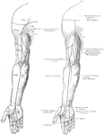
Distribution of cutaneous nerves on the front of the right superior extremity. The figure at the right shows the areas of distribution of the cutaneous nerves. The letters and numerals in the figure at the left indicate the spinal origin of the nerves distributed to each area.

Cutaneous Nerves of the Front of the Arm

Distribution of cutaneous nerves on the front of the right superior extremity. The figure at the right…

The side angle view of a person's head.

Head Profile

The side angle view of a person's head.

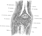
Frontal section of the elbow, viewed from in front.

Frontal Section Through the Elbow

Frontal section of the elbow, viewed from in front.

An illustration of the front of a Pennsylvanian barn.

The Pennsylvanian barn

An illustration of the front of a Pennsylvanian barn.

Superficial muscles of the right thigh, anterior view.

Anterior View of the Superficial Muscles of the Thigh

Superficial muscles of the right thigh, anterior view.
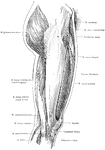
Superficial muscles of the right thigh, lateral view.

Lateral View of the Superficial Muscles of the Thigh

Superficial muscles of the right thigh, lateral view.

Superficial muscles of the right thigh, posterior view.

Posterior View of the Superficial Muscles of the Thigh

Superficial muscles of the right thigh, posterior view.

Superficial muscles of the right leg, anterior view.

Anterior View of the Superficial Muscles of the Leg

Superficial muscles of the right leg, anterior view.

Superficial muscles of the right leg, lateral view.

Lateral View of the Superficial Muscles of the Leg

Superficial muscles of the right leg, lateral view.

Superficial muscles of the right leg, posterior view.

Posterior View of the Superficial Muscles of the Leg

Superficial muscles of the right leg, posterior view.
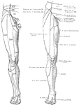
Distribution of cutaneous nerves on the front of the right lower extremity. The figure at the right shows the areas of distribution of the cutaneous nerves. The letters and numeral in the figure at the left indicate spinal origin of the nerves distributed to each area.

Cutaneous Nerves on the Front of the Legs

Distribution of cutaneous nerves on the front of the right lower extremity. The figure at the right…
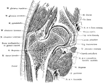
Frontal section through the right hip joint, viewed from in front.

Frontal Section Through Hip Joint

Frontal section through the right hip joint, viewed from in front.
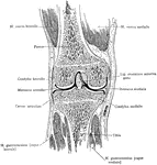
Median frontal section of the right knee, viewed from in front.

Median Frontal Section Through Knee

Median frontal section of the right knee, viewed from in front.

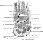
Frontal section of the right ankle and foot. Viewed from in front.

Frontal Section of Foot and Ankle

Frontal section of the right ankle and foot. Viewed from in front.
Fourth cervical vertebra from in front.

Front View of Fourth Cervical

Fourth cervical vertebra from in front.

Fourth cervical vertebra from the side.

Side View of Fourth Cervical

Fourth cervical vertebra from the side.

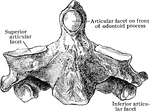
The axis from in front.

Front View of Axis

The axis from in front.

The sacrum, lateral view.

Lateral View of the Sacrum

The sacrum, lateral view.

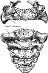
Superior and anterior view of young sacrum of about five years.

Superior and Anterior Surface of Sacrum

Superior and anterior view of young sacrum of about five years.

Anterior and posterior view of adult spine.

Anterior and Posterior Views of Spine

Anterior and posterior view of adult spine.

Lateral view of adult spine.

Lateral View of Spine

Lateral view of adult spine.