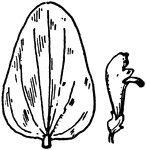
Of the Mint family (Labiatae), the leaf of Scutellaria versicolor.

S. versicolor Skullcap

Of the Mint family (Labiatae), the leaf of Scutellaria versicolor.

Of the Mint family (Labiatae), the leaf of Scutellaria parvula.

S. parvula Skullcap

Of the Mint family (Labiatae), the leaf of Scutellaria parvula.

Of the Mint family (Labiatae), the leaf of Scutellaria canescens.

S. canescens Skullcap

Of the Mint family (Labiatae), the leaf of Scutellaria canescens.

Of the Mint family (Labiatae), the leaf of Scutellaria serrata.

S. serrata Skullcap

Of the Mint family (Labiatae), the leaf of Scutellaria serrata.

Of the Mint family (Labiatae), the leaf of Scutellaria pilosa.

S. pilosa Skullcap

Of the Mint family (Labiatae), the leaf of Scutellaria pilosa.

Of the Mint family (Labiatae), the larger skullcap (Scutellaria integrifolia.

Larger Skullcap

Of the Mint family (Labiatae), the larger skullcap (Scutellaria integrifolia.

The skull of the pterodactyle.

Pterodactyle Skull

The skull of the pterodactyle.

"Fossil skull of the mososaurus." -Taylor, 1904

Mososaurus Skull

"Fossil skull of the mososaurus." -Taylor, 1904

"Skull and antlers of the Irish elk." -Taylor, 1904

Irish Elk Skull

"Skull and antlers of the Irish elk." -Taylor, 1904

Bones of the head.

Bones of the Head

Bones of the head.

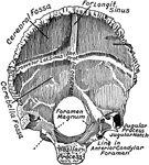
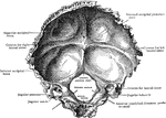
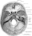
The base of the skull viewed from above. Three Fossae are recognized -the Anterior, Middle, and Posterior Fossae of the Skull.

Base of the Skull

The base of the skull viewed from above. Three Fossae are recognized -the Anterior, Middle, and Posterior…

The skull and spinal canal of a child from behind with the Dura Mater slit open and ribs with the transverse processes of vertebrae dissected out.

The Position of the Spinal Cord and Spinal Nerves in the Spinal Canal

The skull and spinal canal of a child from behind with the Dura Mater slit open and ribs with the transverse…
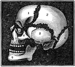
A diagram of the bones of the head. Label: 1, frontal lobe; 2, parietal bone; 3, temporal bone; 4, occipital bone; 5, nasal bone; 6, malar bone; 7, upper jaw; 8, os unguis; 9, lower jaw.

Bones of the Head

A diagram of the bones of the head. Label: 1, frontal lobe; 2, parietal bone; 3, temporal bone; 4, occipital…
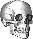
The human skull. Labels: 1, frontal lobe; 2, parietal lobe; 3, temporal lobe; 4, the sphenoid bone; 5, ethmoid bone; 6, superior maxillary (upper jaw) bone; 7, malar bone; 8, lachrymal bone; 9, nasal bone; 10, inferior maxillary (lower jaw) bone.

The Skull

The human skull. Labels: 1, frontal lobe; 2, parietal lobe; 3, temporal lobe; 4, the sphenoid bone;…
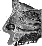
The olfactory system. Labels: a, b, c, d, interior of the nose, which is lined by a mucous membrane; n, the nose; e, the wing of the nose; q, the nose bones; o, the upper lip; g, section of the upper jawbone; h, the upper part of the mouth, or hard palate; m, frontal bone of the skull; k, the ganglion or bulb of the olfactory nerve in the skull, from which are seen the branches of the nerve passing in all direction.

Olfactory System

The olfactory system. Labels: a, b, c, d, interior of the nose, which is lined by a mucous membrane;…

View of the cerebrospinal axis of the nervous system. The right half of the cranium and trunk of the body have been removed by a vertical section; the membranes of the brain and spinal cord have also been removed, and the roots and first part of the fifth and ninth cranial, and of all the spinal nerves of the right side, have been dissected out and laid separately on the wall of the skull and on the several vertebrae opposite to the place of their natural exit from the craniospinal cavity.

Central Nervous System

View of the cerebrospinal axis of the nervous system. The right half of the cranium and trunk of the…

Brain of dog, viewed from above and in profile. F, frontal fissure sometimes termed crucial sulcus, corresponding to the fissure of Rolando in man. S, fissure of Sylvius, around which the four longitudinal convolutions are concentrically arranged; 1, flexion of head on the neck, in the median line; 2, flexion of head on the anterior limb; 5, 6, flexion and extension of posterior limb; 7, 8, 9, contraction of orbicularis oculi, and the facial muscles in general. The unshaded part in that exposed by the opening of the skull.

Brain of a Dog

Brain of dog, viewed from above and in profile. F, frontal fissure sometimes termed crucial sulcus,…
The skull of a newly born child, from above.

Skull of a Newborn

The skull of a newly born child, from above.

Showing the lines which indicate the position of the principal fissures of the brain.

Principle Fissures of the Brain

Showing the lines which indicate the position of the principal fissures of the brain.
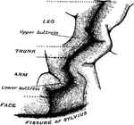
The convolutionary projections of the precentral gyrus, and their relationship to motor areas.

Precentral Gyrus in the Brain

The convolutionary projections of the precentral gyrus, and their relationship to motor areas.
A large moth named for the skull-like image upon its thorax.

Death's Head Moth

A large moth named for the skull-like image upon its thorax.

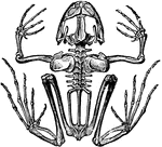
"The skull in reptiles is flat, and the cerebral cavity is not filled with brains. There are no ribs."
Skeleton of a Frog

"The skull in reptiles is flat, and the cerebral cavity is not filled with brains. There are no ribs."

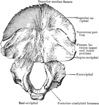
The occipital bone, viewed from above.

Occipital Bone

The occipital bone, viewed from above.

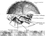
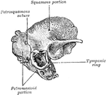
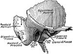
The outer surface of the temporal bone. The dotted lines indicate the lines of suture between squamous, mastoid, or tympanic portions.

Temporal Bone

The outer surface of the temporal bone. The dotted lines indicate the lines of suture between squamous,…
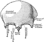
The frontal bone, seen from in front.

Frontal Bone

The frontal bone, seen from in front.

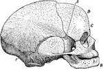
Side view of a fetal skull. The coronal suture extends from the top of the head downwards on either side to the point E; the lambdoidal suture from the back forward, on either side to the point F. The sagittal suture is not shown, but is indicated by the upper margin of the illustration, beginning at the base of the nose, passing backward across the coronal suture (anterior fontanelle), and ending at the lambdoidal suture (posterior fontanelle).

Side View of Fetal Skull

Side view of a fetal skull. The coronal suture extends from the top of the head downwards on either…
Superior maxillary bone and the bony sinuses of the head.

Superior Maxillary Bone and Sinuses

Superior maxillary bone and the bony sinuses of the head.

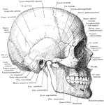
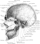
Skull and mandible, lateral view.

Skull and Mandible

Skull and mandible, lateral view.

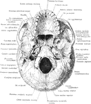
Base of skull.

Base of Skull

Base of skull.

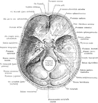
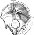
Base of skull from within.

Base of Skull from Within

Base of skull from within.

Median section of the skull and mandible, viewed from the left.

Median Section of the Skull and Mandible

Median section of the skull and mandible, viewed from the left.
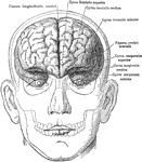
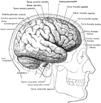
The brain in relation to the skull and face, anterior view.

Brain in Relation to Skull and Face

The brain in relation to the skull and face, anterior view.

The brain in relation to the skull and face, lateral view.

Brain in Relation to Skull and Face

The brain in relation to the skull and face, lateral view.

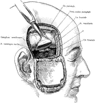
Exposure of the Gasserian ganglion and middle meningeal artery though a flap incision of the scalp and skull. The dura mater and brain are retracted upwards. The (*) points to the Foramen spinosum through which the middle meningeal artery passes as it enters the cranial cavity.

Incision of the Head Showing Gasserian Ganglion

Exposure of the Gasserian ganglion and middle meningeal artery though a flap incision of the scalp and…
Occipital bone, internal surface, from before.

Internal Surface of the Occipital Bone

Occipital bone, internal surface, from before.

Occipital bone at birth.

Occipital Bone at Birth

Occipital bone at birth.

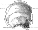
The frontal bone from behind.

Frontal Bone from Behind

The frontal bone from behind.

Right parietal bone, outer surface.

Outer Surface of the Parietal Bone

Right parietal bone, outer surface.

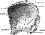
Right parietal bone, inner surface.

Inner Surface of the Parietal Bone

Right parietal bone, inner surface.

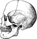
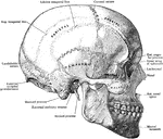
The skull from the side.

Skull from Side

The skull from the side.

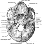
Base of skull from below, the lower jaw is removed.

Base of Skull from Below

Base of skull from below, the lower jaw is removed.

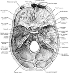
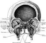
Base of skull from above.

Base of Skull from Above

Base of skull from above.

Front section of skull through plane of outer border of orbits. Arrows pass through communication between atrum and middle meatus.

Frontal Section of Skull Showing Nasal Cavity

Front section of skull through plane of outer border of orbits. Arrows pass through communication between…

Inner aspect of outer wall of right nasal fossa.

Side View of Skull Showing Nasal Cavity

Inner aspect of outer wall of right nasal fossa.

Lateral view of skull with zygomatic arch removed.

Lateral View of Skull

Lateral view of skull with zygomatic arch removed.

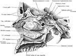
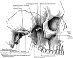
Portion of right half of skull, showing posterior wall of sphenomaxillary fossa. The superior maxilla, ethmoid, and part of malar have been removed.

Skull Showing Posterior Wall of Sphenomaxillary Fossa

Portion of right half of skull, showing posterior wall of sphenomaxillary fossa. The superior maxilla,…

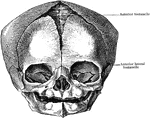
The skull at birth, from before.

Skull at Birth from Before

The skull at birth, from before.

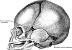
The skull at birth, lateral aspect.

Lateral Aspect of Skull at Birth

The skull at birth, lateral aspect.

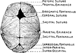
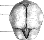
The skull at birth, from above.

Skull at Birth from Above

The skull at birth, from above.

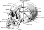
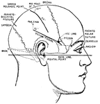
Lateral aspect of the skull, showing the various points.

Skull Showing Various Points

Lateral aspect of the skull, showing the various points.

The inner surface of the occipital bone.

Occipital Bone

The inner surface of the occipital bone.

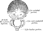
Development of occipital bone. From seven centers.

Development of Occipital Bone

Development of occipital bone. From seven centers.

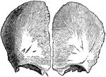
The frontal bone at birth. Developed from two lateral halves.

Frontal Bone at Birth

The frontal bone at birth. Developed from two lateral halves.

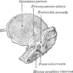
The inner surface of the left temporal bone at birth.

Temporal Bone at Birth

The inner surface of the left temporal bone at birth.

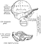
Development of the temporal bone. From ten centers.

Development of Temporal Bone

Development of the temporal bone. From ten centers.

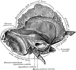
Temporal bone at birth. Outer aspect.

Outer Aspect of Temporal Bone at Birth

Temporal bone at birth. Outer aspect.

Temporal bone at birth. Inner aspect.

Inner Aspect of Temporal Bone at Birth

Temporal bone at birth. Inner aspect.

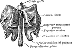
The outer surface of the ethmoid bone.

Outer Surface of Ethmoid Bone

The outer surface of the ethmoid bone.

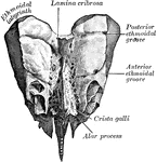
Ethmoid bone from behind.

Ethmoid Bone from Behind

Ethmoid bone from behind.

Ethmoid bone from above.

Ethmoid Bone from Above

Ethmoid bone from above.