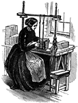
When binding books they are "sewed on a frame, each sheet being attached by a thread to cords across the back. The sewing, though sometimes done by a machine, is cheifly executed by girls."-Lupton

Book Binding

When binding books they are "sewed on a frame, each sheet being attached by a thread to cords across…
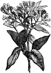
Cloves trees are native to Indonesia. Flower buds from the tree, once dried, are used as a spice in food world wide.

Clove Tree

Cloves trees are native to Indonesia. Flower buds from the tree, once dried, are used as a spice in…

"The modern system of cotton manufacture dates no further back than back 1760. Prior the mechanical inventions of Hargeaves, Arkwright, Crompton and Cartwright, the arts of spinning and weaving were entirely domestic, and the instruments of manipulation much the same as those which had been in use in the East for centuries before." -Lupton

The Spinning Jenny

"The modern system of cotton manufacture dates no further back than back 1760. Prior the mechanical…
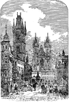
"The capital of Bohemian, a prosperous well-built city near the centre of the kingdom, on both sides of the Moldau, here crossed by seven bridges; 153 miles northwest of Vienna and 75 miles southeast of Dresden, with both of which it is connected b railroad." -Marshall

View in Prague

"The capital of Bohemian, a prosperous well-built city near the centre of the kingdom, on both sides…

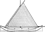
This illustration displays an end view of the proa. A proa is a type of sailing vessel with multi hulls.
End View of Proa

This illustration displays an end view of the proa. A proa is a type of sailing vessel with multi hulls.

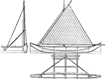
This illustration displays a plan view of the proa. A proa is a type of sailing vessel with multi hulls.
Plan View of Proa

This illustration displays a plan view of the proa. A proa is a type of sailing vessel with multi hulls.

This illustration displays an elevation view of the proa. A proa is a type of sailing vessel with multi hulls.

Elevation View of Proa

This illustration displays an elevation view of the proa. A proa is a type of sailing vessel with multi…

"Prostyle, in architecture, applied to a portico in which the columns stand out quite free from the wall of the building to which it is attached; also applied to a temple or other structure having pillars in front only." -Marshall

Plan view of a Prostyle Temple

"Prostyle, in architecture, applied to a portico in which the columns stand out quite free from the…
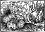
This gigantic flower, one of the marvels of the vegetable world, was discovered in the interior Sumatra by Sir. T Raffles and Dr. Arnold. The whole plant seems to consist of little else beyond the flower and root.

Raffle'sia Flower

This gigantic flower, one of the marvels of the vegetable world, was discovered in the interior Sumatra…
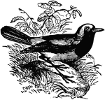
A bird belonging to the family Sylviadae, nearly allied to the nearest, but having a more slender form and a more slender bill. It is found in almost all parts of Britain as a summer bird of passage, and has a soft sweet song.

Redstart Bird

A bird belonging to the family Sylviadae, nearly allied to the nearest, but having a more slender form…
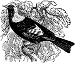
A very beautiful bird of Australia, belonging to the family Meliphagidae or honey-eaters. The color of the plumage is golden yellow and deep velvety black. It was discovered during the regency of George IV., and was named in compliment to him.

Regent Bird

A very beautiful bird of Australia, belonging to the family Meliphagidae or honey-eaters. The color…

The Statue of Liberty in New York is the largest statue in the world, given as a gift from France to celebrate 100 years of United States independence.

Statue of Liberty

The Statue of Liberty in New York is the largest statue in the world, given as a gift from France to…
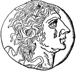
Mithridates VI, king of Pontus in Rome on a silver coin.

Mithridates

Mithridates VI, king of Pontus in Rome on a silver coin.

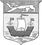
The coat of arms for New Brunswick, a province of Canada.

New Brunswick Coat of Arms

The coat of arms for New Brunswick, a province of Canada.

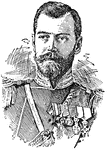
(1868-1918) Nicholas II or Nikolay Alexandrovic Romanov, czar of Russia, king of Poland, and grand duke of Finland.

Nicholas II

(1868-1918) Nicholas II or Nikolay Alexandrovic Romanov, czar of Russia, king of Poland, and grand duke…

A view of Rome including St. Peter's and the Vatican.

View of St. Peter's

A view of Rome including St. Peter's and the Vatican.

A simple king-post roof.

King-post Roof

A simple king-post roof.

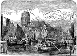
A view of the Netherlands city Rotterdam.

Rotterdam Netherlands

A view of the Netherlands city Rotterdam.

A fish of the carp family, having the back of an olive color; the sides and belly yellow, marked with red; the ventral and anal fins and tail of a deep-red color.

Rudd

A fish of the carp family, having the back of an olive color; the sides and belly yellow, marked with…
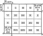
"Here we see 1/2 of 3/4 lb. of candy= 3/8 lb. candy. Use inch, cubes and pennies to show such relations. Let a child have 20 pennies for his money. Let him show 1/2 of 1/2, 1/2 of 1/5, 1/5 of 1/2, 2/5 of 1/2 of his money, etc." -Foster, 1921

Mixed Numbers

"Here we see 1/2 of 3/4 lb. of candy= 3/8 lb. candy. Use inch, cubes and pennies to show such relations.…
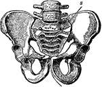
A bony structure located at the bottom of the spine. The human sacrum forms the back part of the pelvis, is roughly triangular in shape, consists of five united vertebrae, and from its solidity it is well adapted to serve as the keystone of the pelvic arch.

Pelvic Bone, Male

A bony structure located at the bottom of the spine. The human sacrum forms the back part of the pelvis,…
Assyrian sculpture of displaying horses, carriage, Assyrians and a lion.

Assyrian Sculpture

Assyrian sculpture of displaying horses, carriage, Assyrians and a lion.

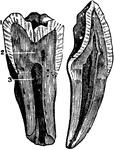
The internal view of a tooth cut through from the top or crown to the tips of the root. Labels: 1, enamel; 2, dentine; 3, pulp

The Structure of a Tooth

The internal view of a tooth cut through from the top or crown to the tips of the root. Labels: 1, enamel;…

Labels: O, esophagus; S, stomach; SI, small intestine; LI, large intestine, Sp spleen; L, liver (raised up); G, gall bladder; Pa, pancreas.

General View of the Alimentary Canal

Labels: O, esophagus; S, stomach; SI, small intestine; LI, large intestine, Sp spleen; L, liver (raised…

Labels: T, thyroid cartilage: C, cricoid cartilage; Tr, trachea; H, hyoid bone; E, epiglottis; I, joint of thyroid cartilage.

Side View of the Larynx

Labels: T, thyroid cartilage: C, cricoid cartilage; Tr, trachea; H, hyoid bone; E, epiglottis; I, joint…
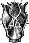
Labels: T, thyroid cartilage: C, cricoid cartilage; Tr, trachea; H, hyoid bone; E, epiglottis; I, joint of thyroid cartilage; Ar, arytenoid cartilages.

Back View of the Larynx

Labels: T, thyroid cartilage: C, cricoid cartilage; Tr, trachea; H, hyoid bone; E, epiglottis; I, joint…
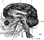
The brain and the cranial nerves seen partly in section and partly in side view. Labels: C, convolutions of the cerebrum; Cbl, cerebellum in section; MD medulla oblongata: Sp N, spinal nerve. The numbers indicate the twelve pairs of cranial nerves in order.

Brain and Cranial Nerves

The brain and the cranial nerves seen partly in section and partly in side view. Labels: C, convolutions…

The chief organs of the body from the side. Labels: a, arch of the aorta or main artery of the trunk; c, large intestine; d, diaphragm; e, throat; g, lung; k, kidney; l, spinal cord within the back bone; m, main artery divided to go to h legs; n, pancreas; o, gullet; s, spleen; tr, wind pipe; t, main vein of body; ap, appendix.

View of Organs from the Side

The chief organs of the body from the side. Labels: a, arch of the aorta or main artery of the trunk;…

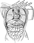
Organs in the body cavity viewed from the front.

View of Organs from the Front

Organs in the body cavity viewed from the front.

Chief veins and arteries of the body. Labels: a, place of the heart; the veins are in the back. On the right side of the picture the veins just under the skin are shown on the other side the deep vessels near the bones. b, vessels to the lungs.

Veins and Arteries of the Body

Chief veins and arteries of the body. Labels: a, place of the heart; the veins are in the back. On the…
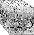
A magnified block of the skin. Labels: a, dead part; d, live part of the epidermis; e, sweat glands; n, nerve endings.

Magnified Image of the Skin

A magnified block of the skin. Labels: a, dead part; d, live part of the epidermis; e, sweat glands;…

The extensor muscles on the back of the forearm. Note the tendons at the wrist.

Muscles on Forearm

The extensor muscles on the back of the forearm. Note the tendons at the wrist.

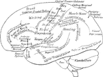
A diagrammatic view displaying the functions of the brain.

Lateral Surface of the Brain

A diagrammatic view displaying the functions of the brain.

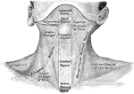
The region of the neck, from in front.

Front View of Neck

The region of the neck, from in front.

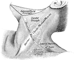
The region of the neck, from the side.

Side View of Neck

The region of the neck, from the side.

A view from behind of a frontal section through the right elbow joint.

Frontal Section Through Elbow Joint

A view from behind of a frontal section through the right elbow joint.

A sagittal section through the left elbow joint of a child. View from the inner side.

Sagittal Section Through Elbow Joint

A sagittal section through the left elbow joint of a child. View from the inner side.
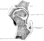
A frontal section through the left hip joint of a boy. Front view.

Frontal Section Through Hip Joint

A frontal section through the left hip joint of a boy. Front view.

A diagram of the relative position of the bone, cartilage, and synovial membrane. Labels: 1,The extremities of two bones to form a joint. 2, The cartilage that covers the end of the bone. 3, The synovial membrane, which covers the cartilage of both bones and is then doubled back from one to the other, represented by the dotted lines.

Position of the Bone, Cartilage, and Synovial Membranes

A diagram of the relative position of the bone, cartilage, and synovial membrane. Labels: 1,The extremities…

The skeleton of a haddock. In some species such as the haddock, there is a modified form of the coracoid bone, free at its lower extremities, which may, perhaps, subserve the purpose of the clavicle of the higher animals.

Skeleton of a Haddock

The skeleton of a haddock. In some species such as the haddock, there is a modified form of the coracoid…

A human skeleton from the back.

Back View of a Human Skeleton

A human skeleton from the back.

A front view of the superficial muscles of the body. Labels: 1, The frontal swells of the occipito-frontalis. 2, The orbicularis palpebrarum. 3, The levator labii superioris alaeque nasi. 4, The zygomaticus major. 5, The zygomaticus minor. 6, The masseter. 7, The orbicularis oris. 8, The depressor labii inferioris. 9, The latysma myoides. 10, The deltoid. 11, The pectoralis major. 12, The latissimus dorsi. 13, The serratus major anticus. 14, The biceps flexor cubiti. 15, The triceps extensor cubiti. 16, The supinator radii longus. 17, The pronator radii teres. 18, The extensor carpi radialis longior. 19, The extensor ossis metacarpi pollicis. 20, The annular ligament. 21, The palmar fascia. 22, The obliquus externus abdominis. 26, The psoas magnus. 27, The abducter longus. 28, The sartorius. 29, The rectus femoris. 30, The vastus externus. 31, vastus internus. 32, The tendo patellae. 33, The gastrocnemius. 34, The tibialis anticus. 35, The tibia. 36, The tendons of the extensor communis.

Front View of the Superficial Muscles of the Body

A front view of the superficial muscles of the body. Labels: 1, The frontal swells of the occipito-frontalis.…
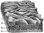
The mucous membrane from the jejunum. Labels: 1, Villi (folds of lining mucous membrane) in miniature. 2, Tubular glands: their orifices. 3, Opening on the free surface of the mucous membrane. 4, Fibrous tissue.

Mucous Membrane from the Jejunum

The mucous membrane from the jejunum. Labels: 1, Villi (folds of lining mucous membrane) in miniature.…

Portion of the mucous membrane from the small intestine, magnified, showing the villi on its free surface, and between them the orifices of the tubular glands. Labels: 1, Portion of a clustered gland. 2, A solitary gland. 3, Fibrous tissue.

A Portion of the Mucous Membrane from the Small Intestine

Portion of the mucous membrane from the small intestine, magnified, showing the villi on its free surface,…

The Annulosa and Mollusca are furnished with a distinct alimentary canal that does not open into the body cavity. In most cases, the digestive canal communicates with the outer world by two-openings- a mouth and an excretory aperture. The annulosa system is demonstrated in the digestive apparatus of a beetle.

The Digestive Apparatus of a Beetle

The Annulosa and Mollusca are furnished with a distinct alimentary canal that does not open into the…

A side view of the lacteal and thoracic duct. Labels: 1, Small intestine. 2, Lacteals. 3, Thoracic duct. 4, Stomach. 5, Colon. 6, Pancreas. 7, Liver. 8, Diaphragm. 9 Heart. 10, Lungs. 11, Large vein into which the thoracic duct opens. 12, Spinal column.

A Side View of the Lacteals and Thoracic Duct

A side view of the lacteal and thoracic duct. Labels: 1, Small intestine. 2, Lacteals. 3, Thoracic duct.…

View of the great lymphatic trunks. Labels: 1, 2 Thoracic duct. 4, The right lymphatic duct. 5, Lymphatics of the thigh. 6, Iliac lymphatics. 7, Lumbar lymphatics. 8, Intercostal lymphatics. a, Descending cava. b, Left innominate vein. c, Right innominate vein. d, Aorta. e, Ascending cava.

View of the Great Lymphatic Trunks

View of the great lymphatic trunks. Labels: 1, 2 Thoracic duct. 4, The right lymphatic duct. 5, Lymphatics…

A side view of the cartilages of the larynx. Labels: *, The front side of the thyroid cartilage. 1, The os hyoides (bone at the base of the tongue). 2, The ligament that connects the hyoid bone and thyroid cartilage. 3, 4, 5, The thyroid cartilage. 6, The cricoid cartilage. 7, The trachea.

A Side View of the Cartilages of the Larynx

A side view of the cartilages of the larynx. Labels: *, The front side of the thyroid cartilage. 1,…

A back view of the cartilages and ligaments of the larynx. Labels: 1, The posterior face of the epiglottis. 3, The os hyoides. 4, The lateral ligaments which connect the os hyoides and thyroid cartilage. 5, The posterior face of the thyroid cartilage. 6, The arytenoid cartilages. 7, The cricoid cartilage. 8, The junction of the cricoid and the arytenoid cartilages. 12, The first ring of the trachea.

A Back View of the Cartilages and Ligaments of the Larynx

A back view of the cartilages and ligaments of the larynx. Labels: 1, The posterior face of the epiglottis.…
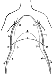
A front view of the chest and abdomen in respiration. Labels: 1, The position of the walls of the chest in inspiration. 2, The position of the diaphragm in inspiration. 3, The position of the walls of the chest in expiration. 4, The position of the diaphragm in expiration. 5, The position of the walls of the abdomen in inspiration. 6, The position of the abdominal walls in expiration.

A Front View of the Chest and Abdomen in Respiration

A front view of the chest and abdomen in respiration. Labels: 1, The position of the walls of the chest…

A side view of the chest and abdomen in respiration. Labels: 1, The cavity of the chest. 2, The cavity of the abdomen. 3, The line of direction for the diaphragm when relaxed in expiration. 4, The line of direction for the diaphragm when contracted in inspiration. 5, 6, The position of the front walls of the chest and abdomen in inspiration. 7, 8, The position of the front walls of the abdomen and chest in expiration.

A Side View of the Chest and Abdomen in Respiration

A side view of the chest and abdomen in respiration. Labels: 1, The cavity of the chest. 2, The cavity…
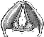
A view of the larynx showing the vocal ligaments. Labels: 1, The anterior edge of the larynx. 4, The posterior face of the thyroid cartilage. 5, The arytenoid cartilages. 6, The vocal ligaments. 7, The origin within the angle of the thyroid cartilage. 9, 10, The glottis.

A View of the Larynx Showing the Vocal Ligaments

A view of the larynx showing the vocal ligaments. Labels: 1, The anterior edge of the larynx. 4, The…

In birds the lungs are confined to the back wall of the chest. They are not separated into lobes, but are oblong and flattened in shape, and connected with a series of air-receptacles scattered through various parts of the body. Shown is the right lung of a goose. Labels: 1, A bronchial tube which divides into two tubes that open into the abdominal air-receptacles (2).

The Right Lung of a Goose

In birds the lungs are confined to the back wall of the chest. They are not separated into lobes, but…
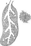
In birds the lungs are confined to the back wall of the chest. They are not separated into lobes, but are oblong and flattened in shape, and connected with a series of air-receptacles scattered through various parts of the body. Shown is a section of a bird lung, magnified. Labels: 1, A bronchial tube that ends in a sac (caeca). 2, Division of the bronchial tubes that branch out among lobules. B, A plexus of capillary vessels.

Section of the Lung of a Bird

In birds the lungs are confined to the back wall of the chest. They are not separated into lobes, but…

A back view of the brain and spinal cord. Labels: 1, The cerebrum. 2, The cerebellum. 3, The spinal cord. 4, Nerves of the face. 5, The brachial plexus of nerves. 6, 7, 8, 9, Nerves of the arm. 10, Nerves that pass under the ribs. 11, The lumbar plexus of nerves. 12, The sacral plexus of nerves. 13, 14, 15, 16, Nerves of the lower limbs.

A Back View of the Brain and Spinal Cord

A back view of the brain and spinal cord. Labels: 1, The cerebrum. 2, The cerebellum. 3, The spinal…
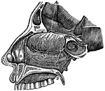
A side view of the passage of the nostrils. 4, The distribution of the first olfactory pair of nerves. 5, The fine divisions of this nerve on the membrane of the nose. 6, A branch of the fifth pair of nerves. 9, Upper jawbone and roof of the mouth.

A Side View of the Passage of the Nostrils

A side view of the passage of the nostrils. 4, The distribution of the first olfactory pair of nerves.…
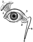
View of the lachrymal gland and nasal duct. Labels: 1, The lachrymal gland. 2, Ducts leading from the lachrymal gland to the upper eyelid. 3, The "tear points". 4, The nasal sac. 5, The termination of the nasal duct.

View of the Lachrymal Gland and Nasal Duct

View of the lachrymal gland and nasal duct. Labels: 1, The lachrymal gland. 2, Ducts leading from the…
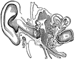
A view of all the parts of the ear, Labels: 1, The tube that leads to the internal ear. 2, The membrana tympani. 3, 4, 5, The bones of the ear. 7, The central part of the labyrinth, names the vestibule. 8, 9, 10, The semicircular canals. 11, 12, The channels of the cochlea. 13, The auditory nerve. 14, The channel from the middle ear to the throat (Eustachian tube). 15, A nerve. 16, A process. 17, The seventh pair of nerves (facial). 18, A process of the temporal bone.

Parts of the Ear

A view of all the parts of the ear, Labels: 1, The tube that leads to the internal ear. 2, The membrana…

"Fault scarps of Orenaug Hill (floating block topography). The view is taken from the top of a cliff in the eastern twin hill." -Walcott, 1901

Fault Scarps

"Fault scarps of Orenaug Hill (floating block topography). The view is taken from the top of a cliff…

"View of the southeastern slope of the eastern twin of Orenaug Hill. Fault scarps bound the hummocky surface of the block on the northeast and west; the courses of the scarps follow the fringes of trees." -Walcott, 1901

Fault Scarp Slope

"View of the southeastern slope of the eastern twin of Orenaug Hill. Fault scarps bound the hummocky…