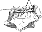
Perpendicular Plate of Ethmoid
Perpendicular plate of ethmoid, shown by removing the right lateral mass.
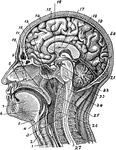
Section of the Head and Neck
Section of head and neck from front to back. Labels: 1, windpipe; 2, larynx; 3, spinal marrow; 4, pharynx;…
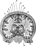
Brain
A cross section of the brain from left to right. Labels: 1, thalamus; 2, skull; 3, cerebral membrane;…
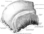
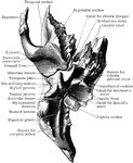
Anterior Half of Section Through Temporal Bone
The anterior half of a vertical transverse section through the left temporal bone.
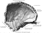
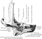
Posterior Half of Section Through Temporal Bone
The posterior half of a vertical transverse section through the left temporal bone.
Horizontal Section of Temporal Bone
Horizontal section through left temporal bone showing lower half of section.
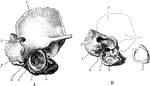
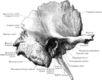
Temporal Bone at Birth
A, The outer surface of the right temporal bone at birth. B, The same with squamozygomatic portion removed.…
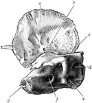
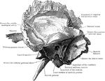
Temporal Bone at Birth
Inner surface of right temporal bone at birth. am squamozygomatic; b, petrosquamosal suture and foramen…
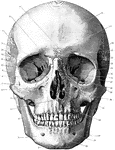
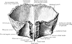
Front of the Skull
Shown is norma frontalis, which refers to the front of the skull. Labels: 1, mental protuberance; 2,…
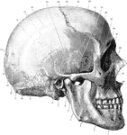
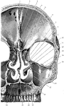
Side of the Skull
Shown is norma lateralis, which refers to the side of the skull. Labels: 1, mental foramen; 2, body…
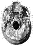
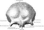
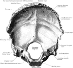
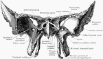
Base of the Skull
Shown is norma basalis, which refers to the base of the cranium. Labels: 1, external occipital crest;…
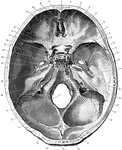
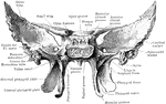
Base of the Skull Seen From Above
Shown is the base of the skull seen from above. Labels: 1, frontal bone; 2, slit for nasal nerve; 3,…
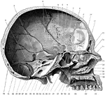
Skull Seen From Side
Shown is the inner aspect of the left half of the skull sagittally divided. Labels: 1, suture between…
Coronal Section of Skull
Shown is a coronal section passing inferiorly through interval between between the first and second…
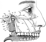
Superior Maxillary Nerve
Scheme of the course and distribution of the superior maxillary nerve. Labels: Rec, recurrent branch…
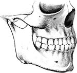
Teeth
To show the relation of the upper to the lower teeth when the mouth is closed. The manner in which a…
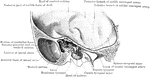
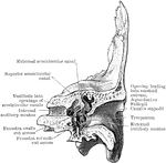
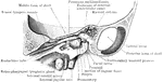
Outer Wall of Tympanic Cavity
View of the outer wall of the middle ear. Section through the left temporal bone of a child to show…
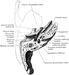
Inner Wall of Tympanic Cavity
View of the inner wall of the middle ear. Section through the left temporal bone of a child to show…
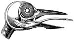
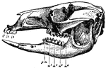
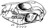
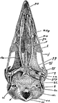
Woodpecker Skull
"Saurognathous skull of woodpecker (Colaptes auratus). v, v, the posterior parts of the abortive vomer;…
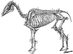
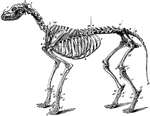
Skeleton of a Horse
The skeleton of a horse. Axial Skeleton. The Skull. Cranial Bones: a, occipital, 1; b, wormian, 1; c,…
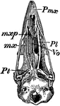
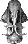
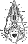
Bird Skull
"Schizognathous skull of common fowl. pmx, premaxilla; mxp, maxillopalatine; mx, maxilla; pl, palatine;…
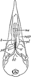
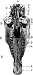
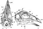
Curlew Skull
"Schizorhinal skull of curlew (top view), showing the long cleft, a, between upper and lower forks of…
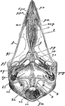
Horse Skull Seen from Above
The skull of a horse seen from above. Labels: I, occipital lobe; II, parietal bone; III, squamosal bone;…
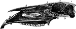
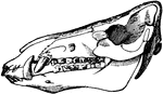
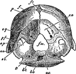
Inferior Aspect of Horse Skull
Inferior aspect of horse's skull, the mandible being removed. Above the line A is the posterior region…
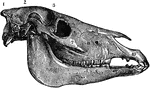
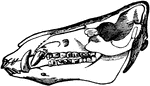
Lateral Aspect of Horse Skull
Lateral aspect of horse's skull. Labels: 1, occipital bone; 2, parietal bone; 3, frontal bone; 9, nasal…
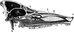
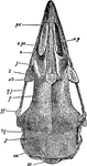
Longitudinal Section of Horse Skull
Longitudinal section of a horse's skull. Labels: 1, supraoccipital bone and crest; 2, parietal bone;…
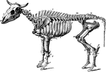
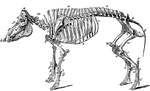
Ox Skeleton
The skeleton of an ox. Axial Skeleton. The skull. Cranial Bones- occipital, 1: b, parietal, 2; a, frontal,…
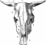
Ox Skull
Skull of an ox, superior aspect. Labels: a, frontal crest; b, lateral crest; c, horn core; d, nasal…
Skeleton of a Hog
Skeleton of the hog. Axial skeleton. The skull. Cranial bones- a, occipital, 1; b, parietal, 2; d, frontal,…
Skull of a Hog
Longitudinal section of a hog's skull. 1, os rostri; 2, maxillary turbinal; 3, frontal turbinal; 4,…
Skeleton of a Dog
The skeleton of the dog. Axial skeleton. The skull. Cranial bones- a, occipital, 1; b, parietal, 2;…
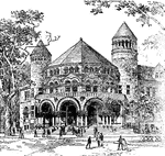
Osborn Hall, Yale University
Many buildings stood on the Old Campus which were removed to make way for the current configuration…
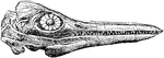
Ichthyosaurus Skull
An illustration of an Ichthyosaurus skull. Ichthyosaurus is an extinct genus of ichthyosaur from the…
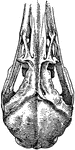
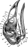
Skull of a Bird
The skull of a bird. Labels: a, inferior aspect, the mandible being removed; b, lateral aspect; px,…
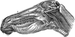
Nostriils of a Horse
Cartilaginous framework of the nostril-seen from above. Labels: a, right alar cartilage; a', left alar…
Head of a Horse
Longitudinal section of the head, showing the pharynx and nasal chamber-the septum nasi being removed.…
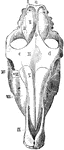
Fowl Skull
The skull of an adult fowl. Here the temporal fossa is bridged over by the junction of the post-frontal…
Roman Leaf Festoon
The Roman Leaf Festoon was hung as a decoration on the friezes of temples, alternating with the real…