Frontal Section Showing Tooth Development
Frontal section showing four earl stages of tooth development.
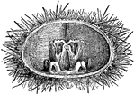
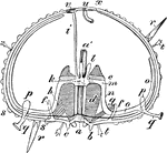
Masticating Apparatus of Echinus Lividus
The organs of mastication of the Sea Urchin. Mastication, or chewing, is the process by which food is…
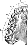
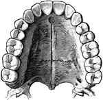
Upper Jaw with Teeth
Right half of upper jaw (from below), with the corresponding teeth. The letters and numbers point to…
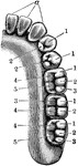
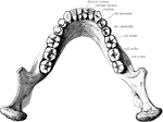
Lower Jaw with Teeth
Right half of lower jaw, with the corresponding teeth. The letters and numbers point to the various…
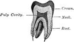
Vertical Section of a Tooth
Vertical section of a tooth in situ. Labels: c, pulp cavity; 1, enamel with radial and concentric markings;…
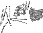
Enamel Prisms
Enamel prisms. A, Fragments and single fibers of the enamel isolated by the action of hydrochloric acid;…
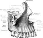

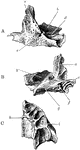
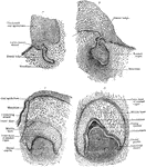
Ossification of Superior Maxilla
Ossification of superior maxilla. A, outer side. B, inner side. C, under side. Labels: a, nasal process;…
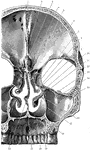
Coronal Section of Skull
Shown is a coronal section passing inferiorly through interval between between the first and second…
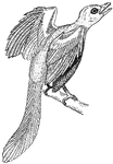
Archaeopteryx
Archaeopteryx, sometimes referred to by its German name Urvogel ("original bird" or "first bird"), is…
Section Through Mouth
Coronal section through the closed mouth. The slit liked character of the vestibule, the manner which…
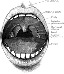
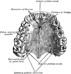
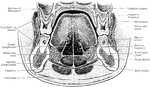
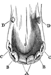
Mouth Showing Palate and Tonsils
Open mouth showing palate and tonsils. It also shows the two palatine arches, and the pharyngeal isthmus…
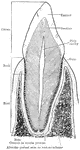
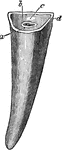
Structure of Canine Tooth
Vertical section of canine tooth to illustrate the various parts and structures.
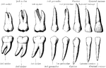
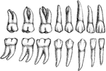
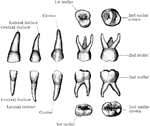
Permanent Teeth
The permanent teeth of the right side, outer or labial aspect. The upper row shows the upper teeth,…
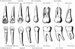
Permanent Teeth
The permanent teeth of the right side, inner of lingual aspect. The upper row shows the upper teeth,…
Jaw Showing Roots of Teeth
Horizontal section through both the upper and lower jaws to show the roots of the teeth. The sections…
Teeth
To show the relation of the upper to the lower teeth when the mouth is closed. The manner in which a…
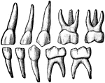
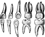
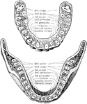
Temporary Teeth
The temporary teeth of the left side. The masticating surfaces of the tow upper molars are shown above.…
Development of a Tooth
Diagram to illustrate the development of a tooth. I. Shows the downgrowth of the dental lamina D.L.…
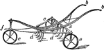
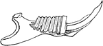
Scarifier
The scarifier is a instrument used for cultivating soil. "a, frame; b, handles; d, teeth; e, wheels;…
Skeleton of a Horse
The skeleton of a horse. Axial Skeleton. The Skull. Cranial Bones: a, occipital, 1; b, wormian, 1; c,…
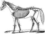
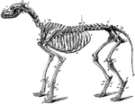
Skeleton of a Horse
The skeleton of a horse. Showing its relation to the contour of the animal, viewed laterally. Labels:…
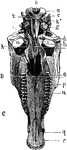
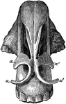
Inferior Aspect of Horse Skull
Inferior aspect of horse's skull, the mandible being removed. Above the line A is the posterior region…
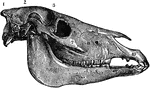
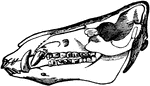
Lateral Aspect of Horse Skull
Lateral aspect of horse's skull. Labels: 1, occipital bone; 2, parietal bone; 3, frontal bone; 9, nasal…
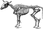
Ox Skeleton
The skeleton of an ox. Axial Skeleton. The skull. Cranial Bones- occipital, 1: b, parietal, 2; a, frontal,…
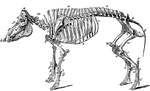
Skeleton of a Hog
Skeleton of the hog. Axial skeleton. The skull. Cranial bones- a, occipital, 1; b, parietal, 2; d, frontal,…
Skeleton of a Dog
The skeleton of the dog. Axial skeleton. The skull. Cranial bones- a, occipital, 1; b, parietal, 2;…
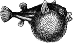
Blowfish
Tetraodontidae is a family of primarily marine and estuarine fish. The family includes many familiar…
Incisor and Canine Horse Teeth
Incisor and canine teeth of a horse. A, front, B, lateral, and C, corner incisor; D, canine teeth.
Horse Incisor Tooth
Incisor tooth of a horse-posterior view. Labels: a, outer layer of enamel; b, inner layer of enamel…
Deadbeat Escapement
The deadbeat has a second face on the pallets, called the 'locking' face, with a curved surface concentric…
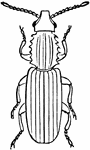
Saw-Toothed Grain Beetle
The Saw-Toothed Grain Beetle (Oryzaephilus surinamensis) is a beetle of the Silvanidae family and is…
Nostriils of a Horse
Cartilaginous framework of the nostril-seen from above. Labels: a, right alar cartilage; a', left alar…
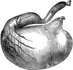
Stomach of a Dog
Stomach of a dog-inflated. Labels: a, cardiac portion; b, pyloric portion; c, esophageal orifice; d,…
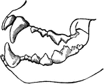
Mandible of a Rabbit
Lateral half of mandible of a rabbi, opened to show the arrangement of rodent teeth.
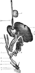
Alimentary Canal of a Bird
Alimentary canal of a bird. Labels: a, ingluvies; b, proventriculus; c, pancreas; d, duodenum; e, liver;…
Rift Saw
"In wood-working, a saw in which the cutting-teeth are placed at the ends of radial arms instead of…
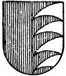
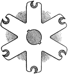
Gules Ordinary
The Gules Ordinary is four wolf's teeth in argent (silver), moving from the sinister (left) side.
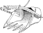
Shovel Bucket
"In railroad construction and canal- or road-work, a large scooping bucket armed with teeth and used…
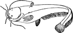
Wels Catfish
The wels catfish is a scaleless fresh and brackish water catfish recognizable by its broad, flat head…
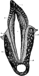
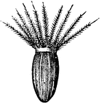
Sea Urchin Section
"Diagram of an Echinus (stripped of its spines). a, mouth; a', gullet; b, teeth; c, lips; d, alveoli;…
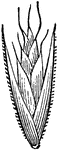
Valerian Seed with Pappus
An illustration of the valerian seed with attached pappus. In a composite flower, Pappus is the part…
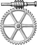
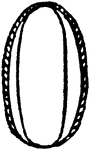
Endless Screw and Wheel
"Endless screw, a mechanical arrangement consisting of a screw the thread of which gears into a wheel…
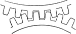
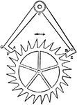
Epicycloidal Teeth
"Epicycloidal teeth, teeth for gearing cut in the form of an epicycloid." -Whitney, 1911
Wood Horsetail
"Equisetum sylvaticum: a, a, sheath crowned with teeth; b, branches; c, c, fruiting spikes. 2. Clypeola,…
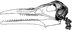
The Restoration of the Hesperornis Regalis
"Hesperornis regalis, (a fossilized restoration) which stood about three feet high, had blunt teeth…
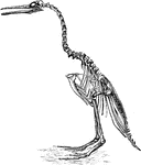
Skeleton Head of a Ichthyornis
"Ichthyornis victor and I. dispar, ...were small forms of about the size of a Partridge, with the habits…
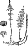
Wild Oat Grass
Wild Oat Grass (Danthonia spicata), also referred to as White Top grass, and Old Fog grass, is common…
Wild Oat Grass
Wild Oat Grass (Danthonia spicata), also referred to as White Top grass, and Old Fog grass, is common…
Wild Oat Grass
Wild Oat Grass (Danthonia spicata), also referred to as White Top grass, and Old Fog grass, is common…