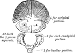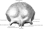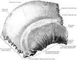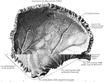
Development of occipital bone. From seven centers.

Development of Occipital Bone

Development of occipital bone. From seven centers.

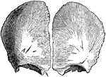
The frontal bone at birth. Developed from two lateral halves.

Frontal Bone at Birth

The frontal bone at birth. Developed from two lateral halves.

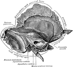
The inner surface of the left temporal bone at birth.

Temporal Bone at Birth

The inner surface of the left temporal bone at birth.

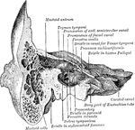
Section through the petrous and mastoid portions of the temporal bone, showing the communication of the cavity of the tympanum with the mastoid antrum.

Section Through Temporal Bone

Section through the petrous and mastoid portions of the temporal bone, showing the communication of…
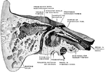
Right temporal bone cut open to show the anterior surface of the petrous portion.

Temporal Bone Cut Open

Right temporal bone cut open to show the anterior surface of the petrous portion.
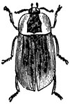
"They introduce themselves under the skin of the carcasses of animals, and devour their flesh to the bone." This one is native to Australia.

Necrodes Lacrymosa

"They introduce themselves under the skin of the carcasses of animals, and devour their flesh to the…
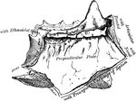
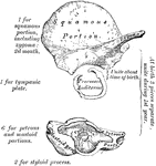
Development of the temporal bone. From ten centers.

Development of Temporal Bone

Development of the temporal bone. From ten centers.

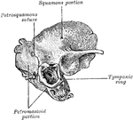
Temporal bone at birth. Outer aspect.

Outer Aspect of Temporal Bone at Birth

Temporal bone at birth. Outer aspect.

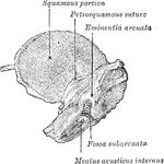
Temporal bone at birth. Inner aspect.

Inner Aspect of Temporal Bone at Birth

Temporal bone at birth. Inner aspect.

The outer surface of the ethmoid bone.

Outer Surface of Ethmoid Bone

The outer surface of the ethmoid bone.

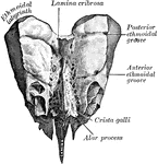
Ethmoid bone from behind.

Ethmoid Bone from Behind

Ethmoid bone from behind.

Ethmoid bone from above.

Ethmoid Bone from Above

Ethmoid bone from above.

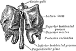
Perpendicular plate of ethmoid, shown by removing the right lateral mass.

Perpendicular Plate of Ethmoid

Perpendicular plate of ethmoid, shown by removing the right lateral mass.

Outer surface of right nasal bone.

Outer Surface of Nasal Bone

Outer surface of right nasal bone.

Inner surface of left nasal bone.

Inner Surface of Nasal Bone

Inner surface of left nasal bone.

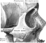
Right malar bone in situ.

Malar Bone

Right malar bone in situ.

Left palate bone. Internal view.

Internal View of Palate

Left palate bone. Internal view.

The turbinated bones are situated one on each side of the outer wall of each nasal fossa. Shown is the internal surface of right turbinated bone.

Internal Surface of Turbinated Bone

The turbinated bones are situated one on each side of the outer wall of each nasal fossa. Shown is the…

The turbinated bones are situated one on each side of the outer wall of each nasal fossa. Shown is the external surface of right turbinated bone.

External Surface of Turbinated Bone

The turbinated bones are situated one on each side of the outer wall of each nasal fossa. Shown is the…

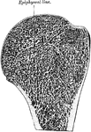
Longitudinal section of head of left humerus.

Longitudinal Section of Head of Humerus

Longitudinal section of head of left humerus.

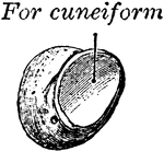
The cuneiform may be distinguished by its pyramidal shape, and by its having an oval, isolated facet for articulation with pisiform bone. It is situated at the upper and inner side of the carpus. Shown is the left cuneiform, showing palmar and lateral surfaces.

Cuneiform Bone

The cuneiform may be distinguished by its pyramidal shape, and by its having an oval, isolated facet…
The pisiform may be known by its small size and by its presenting a single articular facet. It is situated on a plane anterior to the other bones of the carpus; it is spheroidal in form, with its long diameter directed vertically. Shown is the left pisiform, showing posterior and lateral surfaces.

Pisiform Bone

The pisiform may be known by its small size and by its presenting a single articular facet. It is situated…
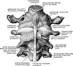
Occipital bone and first three cervical vertebrae with ligaments, from in front.

Occipital Bone and Cervical Vertebrae

Occipital bone and first three cervical vertebrae with ligaments, from in front.

Chain of ossicles and their ligaments, seen from the front in a vertical transverse section of the tympanum.

Ossicles of the Tympanum

Chain of ossicles and their ligaments, seen from the front in a vertical transverse section of the tympanum.

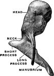
The right malleus viewed from in front.

Malleus From in Front

The right malleus viewed from in front.

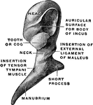
The right malleus viewed from behind.

Malleus From Behind

The right malleus viewed from behind.

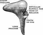
Lateral view of the right incus.

Lateral View of the Incus

Lateral view of the right incus.

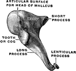
Medial and front view of the right incus.

Medial and Front View of the Incus

Medial and front view of the right incus.

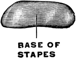
The right stapes viewed from above.

Stapes Viewed from Above

The right stapes viewed from above.

Medial view of the right stapes.

Stapes, Medial View of the

Medial view of the right stapes.

Vertical section of a tooth in situ. Labels: c, pulp cavity; 1, enamel with radial and concentric markings; 2, dentin with tubules and incremental lines; 3, cementum or crusta petrosa; 4, pericemental membrane; 5, bone of mandible.

Vertical Section of a Tooth

Vertical section of a tooth in situ. Labels: c, pulp cavity; 1, enamel with radial and concentric markings;…
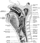
Sagittal median section of the head and neck. The head is thrown backward into complete extension which explains the relations between the lower jaw and the hyoid bone as seen in the figure.

Sagittal Section of the Head and Neck

Sagittal median section of the head and neck. The head is thrown backward into complete extension which…
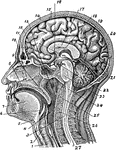
Section of head and neck from front to back. Labels: 1, windpipe; 2, larynx; 3, spinal marrow; 4, pharynx; 5, tongue or hyoid bone; 6, epiglottis; 7, tongue; 8, hard palate; 9, soft palate; 10; bridge of the nose; 11, frontal cavity; 12; sphenoid cavity; 13, nasal cavity; 14, skin of skull; 15, bony skull; 16, hypophisis; 17, corpus callosum; 18, septum lucidum; 19, straight sinus; 20, cerebellum; 21 cerebrum; 22, lobes of medulla; 23, pons varolii; 24, medulla oblongata; 25, zone of the epistropheus; 26, vertebrae; 27, spinal continuation of the vertebrae.

Section of the Head and Neck

Section of head and neck from front to back. Labels: 1, windpipe; 2, larynx; 3, spinal marrow; 4, pharynx;…
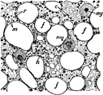
Section of bone marrow. Labels: f, fat vacuole; e, eosinophile cells; ,y, myeloplaxes; r, red corpuscles; m, marrow cells; h, haematoblasts or erythroblasts.

Section of Bone Marrow

Section of bone marrow. Labels: f, fat vacuole; e, eosinophile cells; ,y, myeloplaxes; r, red corpuscles;…
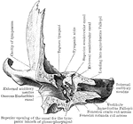
Anterior view of the frontal bone.

Anterior View of Frontal Bone

Anterior view of the frontal bone.

The frontal bone viewed from below.

Frontal Bone Viewed From Below

The frontal bone viewed from below.

The outer side of the right parietal bone.

Outer Surface of the Parietal Bone

The outer side of the right parietal bone.

The inner surface of the right parietal bone.

Inner Surface of the Parietal Bone

The inner surface of the right parietal bone.

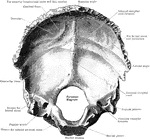
The inner surface of the right occipital bone.

Inner Surface of the Occipital Bone

The inner surface of the right occipital bone.

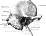
The right temporal bone as seen from the outer side.

Outer Surface of the Temporal Bone

The right temporal bone as seen from the outer side.

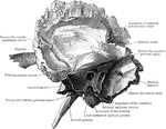
The right temporal bone as seen from the inner side.

Inner Surface of the Temporal Bone

The right temporal bone as seen from the inner side.

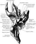
The right temporal bone as seen from below.

Temporal Bone Viewed from Below

The right temporal bone as seen from below.

The anterior half of a vertical transverse section through the left temporal bone.

Anterior Half of Section Through Temporal Bone

The anterior half of a vertical transverse section through the left temporal bone.
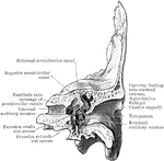
The posterior half of a vertical transverse section through the left temporal bone.

Posterior Half of Section Through Temporal Bone

The posterior half of a vertical transverse section through the left temporal bone.
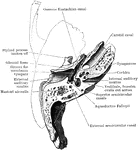
Horizontal section through left temporal bone showing lower half of section.

Horizontal Section of Temporal Bone

Horizontal section through left temporal bone showing lower half of section.

A, The outer surface of the right temporal bone at birth. B, The same with squamozygomatic portion removed. Labels: a, tympanic ring; b, inner wall of tympanum; c, fenestra rotunda; d, foramen ovale; e, mastoid; f, mastoid process; g, mastosquamosal suture, with foramen for transmission of vessels; h, squamozygomatic, removed in figure B to show how its descending process forms the outer wall of the mastoid antrum.

Temporal Bone at Birth

A, The outer surface of the right temporal bone at birth. B, The same with squamozygomatic portion removed.…
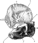
Inner surface of right temporal bone at birth. am squamozygomatic; b, petrosquamosal suture and foramen (just above the end of the lead line; c, subarcuate fossa; d, aquaedutcus vestibuli; e, aquaeductus cochlea; f, internal auditory meatus; g, upper end of carotid canal.

Temporal Bone at Birth

Inner surface of right temporal bone at birth. am squamozygomatic; b, petrosquamosal suture and foramen…

Showing articulation of inferior turbinated bone with ethmoid.

Ethmoid

Showing articulation of inferior turbinated bone with ethmoid.

The outer side (A) and inner side (B) of the right malar bone.

Malar Bone

The outer side (A) and inner side (B) of the right malar bone.

The outer side (A) and inner side (B) of the right nasal bone.

Nasal Bone

The outer side (A) and inner side (B) of the right nasal bone.

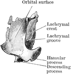
The orbital surface of the right lachrymal bone.

Lachrymal Bone

The orbital surface of the right lachrymal bone.

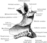
The inner surface (A) and outer surface (B) of the right inferior turbinated bone.

Inferior Turbinated Bone

The inner surface (A) and outer surface (B) of the right inferior turbinated bone.
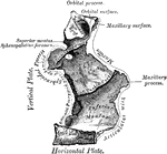
Right palate bone seen from outer side.

Outer Side of Palate Bone

Right palate bone seen from outer side.

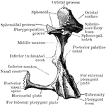
Right palate bone seen from inner side.

Inner Side of Palate Bone

Right palate bone seen from inner side.

Right palate bone as seen from behind.

Palate Bone Viewed from Behind

Right palate bone as seen from behind.

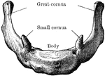
The hyoid bone as seen from in front.

Hyoid Bone

The hyoid bone as seen from in front.

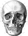
Shown is norma frontalis, which refers to the front of the skull. Labels: 1, mental protuberance; 2, body of lower jaw; 3, ramus of lower jaw; 4, anterior nasal spine; 5, canine fossa; 6, infraorbital canal; 7, malar canal; 8 orbital surface of superior maxilla; 9, temporal fossa; 10, os planum of ethmoid; 11, sphenoidal fissure; 12, lachrymal bone and groove; 13, optic foramen; 14, orbital foramina; 15, temporal ridge; 16, supraorbital notch; 17, glabella; 18, frontal eminence; 19, superciliary ridge; 20, parietal bone; 21, front-nasal suture; 22, pterion; 23, great wing of sphenoid; 24, orbital surface of great wing of sphenoid; 25, squamous temporal; 26, left nasal bone; 27, malar bone; 28, sphenomaxillary fissure; 29, zygomatic arch; 30, anterior nasal aperture, displaying nasal septum and inferior and middle turbinated bones; 31, mastoid process; 32, incisor fossa; 33, angle of jaw; 34, mental foramen; 35, symphysis.

Front of the Skull

Shown is norma frontalis, which refers to the front of the skull. Labels: 1, mental protuberance; 2,…
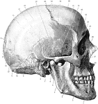
Shown is norma lateralis, which refers to the side of the skull. Labels: 1, mental foramen; 2, body of lower jaw; 3, superior maxillary; 4, ramus of lower jaw; 5, zygomatic arch; 6, styloid process; 7, external auditory meatus; 8, mastoid process; 9, asterion; 10, superior curved line of occipital bone; 11, external occipital protuberance; 12, lambdoid suture; 13, occipital bone; 14, lambda; 15, obelion placed between the two parietal foramina; 16, parietal bone; 17, lower temporal ridge; 18, upper temporal ridge; 19, squamous part of temporal bone; 20, bregma; 21, coronal suture; 22, stephanion; 23, frontal bone; 24, pterion; 25, temporal fossa; 26, great wing of sphenoid; 27, malar bone; 28, malar canal; 29, lachrymal bone; 30, nasal bone; 31, infraorbital canal; 32, anterior nasal aperture.

Side of the Skull

Shown is norma lateralis, which refers to the side of the skull. Labels: 1, mental foramen; 2, body…
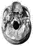
Shown is norma basalis, which refers to the base of the cranium. Labels: 1, external occipital crest; 2, superior curved line of the occipital bone; 3, foramen magnum; 4, occipital condyle; 5, digastric groove; 6, mastoid process; 7, external auditory meatus; 8, styloid process; 9, glenoid fossa; 10, foramen spinosum; 11, sphenoidal spine; 12, foramen ovale; 13, external pterygoid plate; 14, hamular process of internal pterygoid plate; 15, nasal septum; 16, posterior nasal spine; 17, horizontal plate of palate bone; 18, palatal process of superior maxilla; 19, anterior palatine canal; 20, intermaxillary suture; 21, posterior palatine canal; 22, malar process of superior maxilla; 23, spheno-maxillary fissure; 24, zygomatic fossa; 25, zygomatic arch; 26, posterior openings of left nasal fossa; 27, pterygoid fossa; 28, scaphoid fossa; 29, foramen lacerum medium; 30, opening of osseous Eustachian canal; 31, carotid canal; 32, jugular fossa; 33, stylo-mastoid foramen; 34, jugular process of the occipital bone; 35, groove for occipital artery; 36, mastoid foramen; 37, posterior condylic foramen; 38, inferior curved line of occipital bone; 39, external occipital protuberance.

Base of the Skull

Shown is norma basalis, which refers to the base of the cranium. Labels: 1, external occipital crest;…
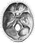
Shown is the base of the skull seen from above. Labels: 1, frontal bone; 2, slit for nasal nerve; 3, anterior ethmoidal foramen; 4, posterior ethmoidal foramen; 5, optic foramen; 6, foramen for internal carotid artery formed by anterior and middle clinoid processes; 7, lesser wing of sphenoid; 8, anterior clinoid process, in this case united on its inner side to the middle clinoid process; 9, posterior clinoid process; 10, foramen ovale; 11, groove for middle meningeal artery; 12, foramen spinosum; 13, Hiatus Fallopii; 14, line of petro-squamosal suture; 15, internal auditory meatus; 16, groove for superior petrosal sinus; 17, groove for sigmoid part of lateral sinus; 18, jugular foramen; 19, anterior condylic foramen; 20, groove for lateral sinus; 21, internal occipital protuberance; 22, ridge for attachment of falx cerebri; 23, fossa for the lodgment of the occipital lobe of the cerebrum; 24, ridge for attachment of the falx cerebelli; 25, fossa for the lodgment of the left cerebellar hemisphere; 26, foramen magnum; 27, groove for the sigmoid sinus turning into the jugular foramen; 28, groove for the inferior petrosal sinus running along the line of the suture between the petrous temporal and the basioccipital; 29, depression for the Gasserian ganglion; 30, middle cranial fossa for lodgment of temporal lobe of cerebrum; 31, foramen lacerum medium; 32, carotid groove; 33, dorsum sellae of sphenoid; 34, leads into foramen rotundum; 35, pituitary fossa; 36, olivary eminence of sphenoid; 37, anterior cranial fossa for lodgment of frontal lobes of cerebrum; 38, cribriform plate of ethmoid; 39, crista galli of ethmoid; 40, foramen caecum; 41, crest for attachment of falx cerebri.

Base of the Skull Seen From Above

Shown is the base of the skull seen from above. Labels: 1, frontal bone; 2, slit for nasal nerve; 3,…