Clipart tagged: ‘abdomen’
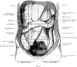
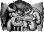
Abdomen Laid Open After Removal of Jejunum and Ileum
The abdomen viscera after the removal of the jejunum and ileum. The transverse colon is much more regular…
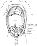
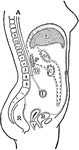
Abdomen of Fetus
The abdominal and thoracic viscera of a five months fetus. The large liver and large size if its left…
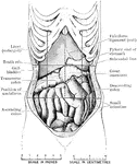
Abdomen Showing Displacement Caused by Corset
Abdomen of female showing displacement resulting from tight lacing. The liver is much enlarged, and…

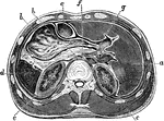
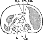
Horizontal Section Through Abdomen
Horizontal section through upper part of abdomen. Labels: a, liver; b, stomach; c, transverse colon;…
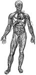
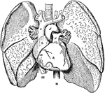
Principal Organs of the Thorax and Abdomen
"The principal muscles are seen on the left, and superficial veins on the right." — Blaisedell, 1904
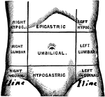
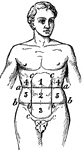
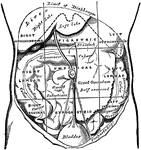
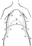
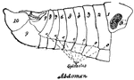
Regions of the Abdomen
Regions of the abdomen. The abdomen is divided into 9 regions by two horizontal planes, one at the level…
Transverse Section of Abdomen
Diagrammatic transverse section of abdomen, to show the peritoneum on transverse tracing. A, at level…
Abdominal
"Pertaining to the abdomen or belly; situated in or on the abdomen: as abdominal ventral fins."-Wright,
Abdominal
"In human anatomy, certain regions into which the abdomen is arbitrarily divided for the purpose of…
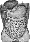
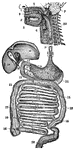
Abdominal Organs
Abdominal organs. Labels: 1, liver turned up; 2, gall bladder; 3, stomach; 4, large intestine; 5, small…
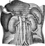
Abdominal Organs
Abdominal organs. Labels: 1, liver turned up; 2, gall bladder; 3, right kidney; 4, spleen; 5, left kidney.
Regions of the Abdomen and their Contents
Regions of the abdomen and their contents (edge of costal cartilages in dotted outline). "For convenience…
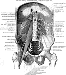
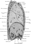
Muscles of the Abdominal Wall
View of the posterior abdominal wall to show the muscles and the nerves of the lumbo sacral plexus.
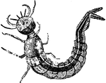
Water Beetle Larva
Water beetles carry air bubbles under their abdomens, which provides an air supply. Water beetle larvae…
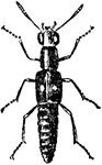
Brachelytra
"Readily distinguished from the other groups of beetles by having the elytra much shorter then the abdomen,…
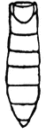
Monarch Butterfly
The abdomen of the monarch. This common butterfly is of a brown color, with black veins and wing borders.
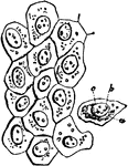
Cells from the Abdominal Lining
Flat cells from the surface of the lining membrane of the abdomen (peritoneum). Labels: a, cell-body;…
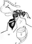
Female Chalcid Wasp
The female chalcid wasp (Eurytoma prunicola) is a parasitoid in the family Chalcididae.
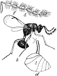
Male Chalcid Wasp
The male chalcid wasp (Eurytoma prunicola) is a parasitoid in the family Chalcididae.
A Front View of the Chest and Abdomen in Respiration
A front view of the chest and abdomen in respiration. Labels: 1, The position of the walls of the chest…
A Side View of the Chest and Abdomen in Respiration
A side view of the chest and abdomen in respiration. Labels: 1, The cavity of the chest. 2, The cavity…
Diaphragm
View of the diaphragm; 1, cavity of the thorax; 2, diaphragm separating the cavity of the thorax from…
Digestive System
A diagram of the organs of digestion. Labels:1, The upper jaw. 2, The lower jaw. 3, The tongue. 4, The…
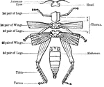
External Anatomy of an Insect Skeleton
"Anatomy of the external skeleton of an insect" — Goodrich, 1859
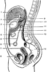
The Peritoneum
The peritoneum is a large serous membrane, which forms in the male a closed sac, the parietal layer…
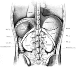
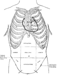
Diagram of Thoracic and Abdominal Regions
A diagram of the thoracic and abdominal regions. Labels: A, aortic valve; M, mitral valve; p, pulmonary…

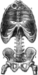
Thorax and Abdomen
"Thorax and abdomen. 1, 1, 1, 1. Muscles of the chest. 2, 2, 2, 2. Ribs. 3, 3, 3. Upper, middle and…
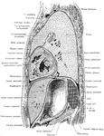
Side View of the Thorax and Part of the Abdomen
Lateral, sagittal section through the left thorax and upper portion of abdomen, viewed from the left.…
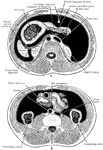
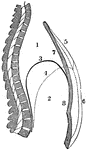
Horizontal Section Through Trunk
Diagram of horizontal section through upper part of 1st lumbar vertebra. The fine dots represent the…
Side View of the Trunk
Sagittal section through the trunk, 6 cm to the right of the median plane, viewed from the right. Note…
Transverse Section Through
Transverse section through the abdomen, opposite the second lumbar vertebra.
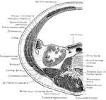
Vertical Median Section of the Trunk
Diagram of vertical median section of abdomen. The fine dots represent the great sac of the peritoneum,…