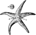
"Antambulacral surface of Asterias rubens. a, madreporite; a', the same magnified; b, anus." — Encyclopedia Britannica, 1893

Asterius Rubens

"Antambulacral surface of Asterias rubens. a, madreporite; a', the same magnified; b, anus." —…
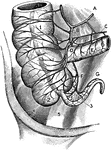
The arterial blood supply of the anterior (ventral) surface of the cecum and appendix. Labels: A, ileocolic artery; B, cecal appendicular artery; D, anterior cecal artery; F and G, Appendicular artery. C, Iliac artery; 1, Right colon; 2, External sacculus of cecum ( to right of taenium coli); 3, Appendix; 4, Iliac muscle; 5, Psoas muscle.

Arterial Blood Supply of the Cecum and Appendix

The arterial blood supply of the anterior (ventral) surface of the cecum and appendix. Labels: A, ileocolic…

A diagram of the digestive system.

Digestive System

A diagram of the digestive system.

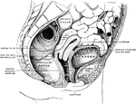
Sagittal section in the median line of the female pelvis.

female Pelvis

Sagittal section in the median line of the female pelvis.

"Galeodes sp., one of the solifugae. Dorsal view. I to VI, Bases of the prosomatic appendages. o, Eyes. a, Lateral region of the cephalic plate to which the first pair of appendages are articulated. b, Cephalic plate with median eye. c, Dorsal element of somites bearing third and fourth pairs of appendages. d, Second plate of the prosoma with fifth pair of appendages. e, Third or hindermost plate of the prosoma beneath which the sixth pair of legs is articulated. 1, 2, 9, 10, First, second, ninth and tenth somites of the opisthosoma. an, Anus." — The Encyclopedia Britannica, 1910

Galeodes

"Galeodes sp., one of the solifugae. Dorsal view. I to VI, Bases of the prosomatic appendages. o, Eyes.…

"Garypus litoralis, one of the Pseudoscorpiones. Ventral view. I to VI, Prosomatic appendages. o, Sterno-coxal process of the basal segment of the second appendage. 1, Sternite of the genital or first opisthosomatic somite; the prae-genital somite, though represented by a tergum, has no seperate sternal plate. 2 and 3, Sternites of the second and third somites of the opishosoma, each showing a tracheal stigma. 10 and 11, Sternites of the tenth and eleventh somites of the opisthosoma. an, Anus. " — The Encyclopedia Britannica, 1910

Garypus Litoralis

"Garypus litoralis, one of the Pseudoscorpiones. Ventral view. I to VI, Prosomatic appendages. o, Sterno-coxal…

"Garypus litoralis, one of the Pseudoscorpiones. Dorsal view. I to VI, The prosomatic appendages. o, Eyes. Prae-gen, Prae-genital somite. 1, Tergite of the genital or first opisthosomatic somite. 10, Tergite of the tenth somite of the opisthosoma. 11, The evanescent eleventh somite of the opisthosoma. an, Anus." — The Encyclopedia Britannica, 1910

Garypus Litoralis

"Garypus litoralis, one of the Pseudoscorpiones. Dorsal view. I to VI, The prosomatic appendages. o,…

"Garypus litoralis, one of the Pseudoscopions. Lateral view. I to VI, Basal segments of the six prosomatic appendages. o, Eyes. prae-gen, Tergite of the prae-genital somite. I, Genital or first opisthosomatic. 2, 3, 10, The second, third and tenth somites of the opisthosoma. 11, The minute eleventh somite. an, The anus." — The Encyclopedia Britannica, 1910

Garypus Litoralis

"Garypus litoralis, one of the Pseudoscopions. Lateral view. I to VI, Basal segments of the six prosomatic…
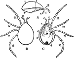
"Holothyrus nitidissimus, one of the Acari; ater Thorell. A, Lateral view with appendages III to VI removed, I, plate covering the whole dorsal area, representing the fused tergal sclerites of the prosoma and opisthosoma; 2, similarly-formed ventral plate; 3, tracheal stigma. B, dorsal view of the same animal; II to VI, 2nd to 6th pairs of appendages. The 1st pair of appendages both in this and in C are retracted. C, Ventral view of the same; II to VI as in B; a, genital orifice; b, anus; c, united basal segments of the second pair of appendages; d, basal segment of the 6th prosomatic appendage of the right side. The rest of the appendage, as also of app. III, IV and V, has been cut away." — The Encyclopedia Britannica, 1910

Holothyrus Nitidissimus

"Holothyrus nitidissimus, one of the Acari; ater Thorell. A, Lateral view with appendages III to VI…

"Liphistus desultor, Ventral view with the prosomatic appendages cut short expecting the chelicerae (1) whose sharp retroverts are seen. Between the bases of the prosomatic limbs an anterior and a posterior sternal plate (black) are seen. I, The sternum of the first opisthosomatic or genital somite covering the genital aperture and the first pair of lungsacs. In front of it the narrow waist is formed by the soft sternal area of the praegenital somite. 2, the sternite of the second opisthosomatic somite covering the posterior pair of lung-sacs; 3 and 4, the spinning appendages (limbs) of the opisthosoma; a, inner, b, outer ramus of the appendage; II, sternite of the eleventh somite of the opisthosoma: in front of it other rudimentary sternites; an, anus." — The Encyclopedia Britannica, 1910

Liphistius Desultor

"Liphistus desultor, Ventral view with the prosomatic appendages cut short expecting the chelicerae…
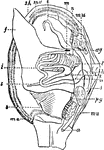
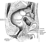
Sagittal section in the median line of the male pelvis.

Male Pelvis

Sagittal section in the median line of the male pelvis.

"Anatomy of an Acephalous Mollusc (Mactra): s, stomach; ii, intestine; ag, anterior ganglions; pg, posterior ganglions; mn, muscles; a, anus; h, heart; l, liver; f, foot; sh, shell; ma, mantle; b, branchiae; t, tentacula; r, oral, or respiratory syphon; t, anal syphon." — Chambers, 1881

Mollusc Anatomy

"Anatomy of an Acephalous Mollusc (Mactra): s, stomach; ii, intestine; ag, anterior ganglions; pg, posterior…
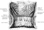
The interior of the anal canal and lower part of the rectum. Showing the column of Morgagni and the anal valves between their lower ends. The columns were more numerous in this specimen than usual.

Rectum and Anal Canal

The interior of the anal canal and lower part of the rectum. Showing the column of Morgagni and the…

The anal canal and rectum in a fetus. A, aged 4 to 5 months; B, 6 moths; C, 9 months. In each he anal canal is distinctly marked off from the rectum proper; the column of Morgagni and the rectal valve are distinct.

Rectum and Anal Canal in Fetus

The anal canal and rectum in a fetus. A, aged 4 to 5 months; B, 6 moths; C, 9 months. In each he anal…

The blood vessels of the rectum and anus, showing the distribution and anastomosis on the posterior surface near the termination of the gut.

Blood Vessels of the Rectum and Anus

The blood vessels of the rectum and anus, showing the distribution and anastomosis on the posterior…

Inner wall of the lower end of the rectum and anus. On the right the mucous membrane has been removed to show the dilation of the veins and how they pass through the muscular wall to anastomose with the external hemorrhoidal plexus.

Inner Wall of the Rectum and Anus

Inner wall of the lower end of the rectum and anus. On the right the mucous membrane has been removed…

"Anatomy of the Snail: a, the mouth; bb, foot; c, anus; dd, lung; e, stomach, covered above by the salivary glands; ff, intestine; g, liver; h, heart; i, aorta; j, gastric artery; l, hepatic artery; k, artery of the foot; mm, abdominal cavity, supplying the place of a cenous sinus; nn, irregular canal in communication with the abdominal cavaty, and carrying the blood to the lung; oo, vessel carrying the blood from the lung to the heart." — Chambers, 1881

Snail Anatomy

"Anatomy of the Snail: a, the mouth; bb, foot; c, anus; dd, lung; e, stomach, covered above by the salivary…

This diagram represents the vertical section through an arm and an interradis of a starfish. a, anus; amp., ampulla; c.b., circular blood vessel; c.w., circular water canal; co., caelom; co.e., caelomic epithelium; d.b., dermal branchae; e, position of the eyespot; ect., ectoderm; ent, entoderm; f, ambulacral foot; g, ambulacral foot; g, ambulacral groove; h, hepatic caeca or liver; i, insestine; i.c., intestinal caeca; mes, mesoderm; mo., mouth; m.p., madreporic body; n.r., nerve ring; os., ossicles in mesoderm; r.n., radial nerve band; r.b., radial blood vessel; r.p., reproductive pore; r.w., radial water canal; s.c., stone canal; sp., spines; z, lacunar spaces in the mesoderm.

Starfish

This diagram represents the vertical section through an arm and an interradis of a starfish. a, anus;…