An oblique fracture of the shaft of the tibia.

Fractured Tibia

An oblique fracture of the shaft of the tibia.

Frontal bone of the human skull, outer surface. The frontal bone forms the forehead, roof of the orbital cavities, and the nasal cavity. Labels: 1, frontal eminence; 7, roof of orbital cavitiy; 10, orbital arch.

Frontal Bone of the Human Skull

Frontal bone of the human skull, outer surface. The frontal bone forms the forehead, roof of the orbital…
The skeletal structure of a gorilla.

Gorilla Skeleton

The skeletal structure of a gorilla.

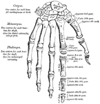
A plan of the development of the bones of the hand.

Hand Development

A plan of the development of the bones of the hand.

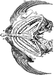
An illustration of a sparrow-hawk's skeleton.

Skeleton of a Sparrowhawk

An illustration of a sparrow-hawk's skeleton.

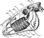
"Side view of skull of horse, with the bone removed so as to expose the whole of the teeth. PMx, premaxilla; Mx, maxilla; Na, nasal bone; Ma, malar bone; L, lacrymal bone; Fr, frontal bone; Sq, squamosal bone; Pa, parietal bone; oc, occipital condyle; pp, paroccipital process; i, the three incisor teeth; c, the canine tooth; pm1, the situation of the rudimentary first premolar, which has been lost in the lower, but is present in the upper jaw; pm2, pm3, and pm4, the three fully-developed premolar teeth; m, the three true molar teeth." — Encyclopedia Britannica, 1893

Horse Skull

"Side view of skull of horse, with the bone removed so as to expose the whole of the teeth. PMx, premaxilla;…

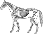
"Skeleton of the horse." — Davison, 1906

Skeleton of a Horse

"Skeleton of the horse." — Davison, 1906

"Side views of skull and hyoid bone of Howling Money." — Encyclopedia Britanica, 1893

Howling Monkey Skull

"Side views of skull and hyoid bone of Howling Money." — Encyclopedia Britanica, 1893

This illustration shows a human leg (front view), and comparative diagrams showing modifications of the leg:
1. Human, 2. Ox, 3. Horse, 4. Bird, 5. Frog, 6. Crocodile, 7. Seal, 8. Dog, 9. Bear
P. Pelvis, FE. Femur, TI. Tibia, FI. Fibula, TA. Tarsus, MT. Metatarsus, PH. Phalanges, OC. Os Calcis.

Human Leg (Front View), and Comparative Diagrams showing Modifications of the Leg

This illustration shows a human leg (front view), and comparative diagrams showing modifications of…

A human skeleton.

Human skeleton

A human skeleton.

"The <em>humerus</em>, a long, hollow bone, rests against a shallow socket on the shoulder blade. It is joined at the elbow to the bones of the forearm." &mdash; Blaisedell, 1904

Humerus

"The humerus, a long, hollow bone, rests against a shallow socket on the shoulder blade. It…

"The humerus, which moves freely by a globular head upon the scapula, forming the shoulder-joint." &mdash; Chambers' Encyclopedia, 1875

Humerus

"The humerus, which moves freely by a globular head upon the scapula, forming the shoulder-joint." —…

The human humerus bone, the longest and largest bone of the upper leg. Labels: a, rounded head; gt, greater tuberosity; lt, lesser tuberosity; b, groove for attachment of biceps tendon.

The Human Humerus

The human humerus bone, the longest and largest bone of the upper leg. Labels: a, rounded head; gt,…
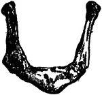
"Hyoid bone of the Perch." &mdash; Encyclopedia Britannica, 1893

Hyoid Bone

"Hyoid bone of the Perch." — Encyclopedia Britannica, 1893

The hyoid, os hyoides, or tongue bone, is an isolated, U-shaped bone lying in front of the throat, just above the Adam's apple; it supports the tongue, and give attachment to some of its numerous muscles.

Human Hyoid Bone

The hyoid, os hyoides, or tongue bone, is an isolated, U-shaped bone lying in front of the throat, just…
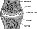
A simple complete joint, one type of movable articulation. The synovial membrane is represented by dotted lines. They have a secreting membrane placed between their opposing surfaces, which keeps them well lubricated and capable of free movement one upon the other.

A Simple Complete Joint

A simple complete joint, one type of movable articulation. The synovial membrane is represented by dotted…
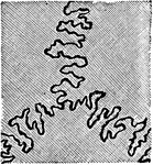
A toothed, or dentated suture. This is one type of immovable articulation. It is found in the union of the cranial bones of the skull, such as the suture between the frontal and parietal bones.

Human Joint, Dentated Suture

A toothed, or dentated suture. This is one type of immovable articulation. It is found in the union…

"Showing how the Ends of the Bones are shaped to form the Elbow Joint. The cut ends of a few ligaments are seen." — Blaisedell, 1904

Elbow Joint

"Showing how the Ends of the Bones are shaped to form the Elbow Joint. The cut ends of a few ligaments…

A mixed articulation (slightly movable). In this form, the bony surfaces are usually joined together by broad, flattened disks of fibro-cartilage, as in the articulations between the bodies of the vertebrae. Labels: a, b, disk of fibro-cartilage; c, articular cartilage; d, bone.

Human Joint, Mixed Articulation

A mixed articulation (slightly movable). In this form, the bony surfaces are usually joined together…

This illustration shows a section on the knee (A, Femur; B, Tibia; C, Patella; D, Synovial sac; E, bursæ). The knee is a hinge-joint, and the bones entering into its formation are the lower end of the femur, the upper end of the tibia, and the posterior surface of the patella (knee-cap).

Section of the Knee

This illustration shows a section on the knee (A, Femur; B, Tibia; C, Patella; D, Synovial sac; E, bursæ).…

Lachrymal Bone. The lachrymal are the smallest and most fragile bones fo the face. They are situated at the front part of the inner wall of the orbit.

Human Lachrymal Facial Bone

Lachrymal Bone. The lachrymal are the smallest and most fragile bones fo the face. They are situated…
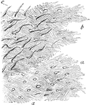
Lamellae torn off from a decalcified human parietal bone at some depth from the surface. Labels: a, lamellae, showing reticular fibers; b, darker part, where several lamellae are superposed; perforating fibers. Apertures through which perforating fibers has passed, are seen especially in the lower part of the figure.

Lamellae

Lamellae torn off from a decalcified human parietal bone at some depth from the surface. Labels: a,…
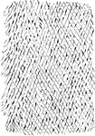
Shown is a thin layer peeled off from a soft bone, which is intended to represent the reticular structure of a lamella.

Reticular Structure of a Lamellae

Shown is a thin layer peeled off from a soft bone, which is intended to represent the reticular structure…

"Bones of the leg. a, femur; b, tibia; c, fibula; d, tarsal bones; e, metatarsal bones; f, phalanges; g, patella." &mdash;Martin, 1917

Leg Bones

"Bones of the leg. a, femur; b, tibia; c, fibula; d, tarsal bones; e, metatarsal bones; f, phalanges;…

"Little openings (Haversian canals) are seen, and around them are arranged rings of bone with little dark spaces (<em>lacunae</em>), from which branch out fine dark lines (<em>analiculi</em>)." &mdash; Blaisedell, 1904

Cross-section from a shaft of a long bone

"Little openings (Haversian canals) are seen, and around them are arranged rings of bone with little…
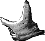
Malar (cheek) bone. The malar bones form the prominence of the cheek, and part of the outer wall and floor of the orbit.

Human Malar (Cheek) Bone

Malar (cheek) bone. The malar bones form the prominence of the cheek, and part of the outer wall and…
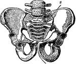
A bony structure located at the bottom of the spine. The human sacrum forms the back part of the pelvis, is roughly triangular in shape, consists of five united vertebrae, and from its solidity it is well adapted to serve as the keystone of the pelvic arch.

Pelvic Bone, Male

A bony structure located at the bottom of the spine. The human sacrum forms the back part of the pelvis,…
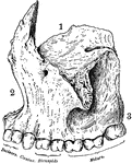
Superior maxillary bone. With it's fellow on the opposite side, it forms the whole of the upper jaw. Each bone assists in forming part of the floor of the orbit, the floor and outer wall of the nasal fossae, and the greater part of the roof of the mouth. Labels: 1, orbital surface; 2, facial surface; 3, alveolar process.

Human Maxillary (Upper Jaw) Bone

Superior maxillary bone. With it's fellow on the opposite side, it forms the whole of the upper jaw.…

Inferior Maxillary Bone (lower jaw). It is the largest and strongest bone in the face and serves for the reception of the lower teeth.

Human Maxillary (Upper Jaw) Bone

Inferior Maxillary Bone (lower jaw). It is the largest and strongest bone in the face and serves for…

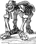
The fossilized skeleton of a Megatherium.

Fossil Megatherium

The fossilized skeleton of a Megatherium.

The first metatarsal of the left foot.

Metatarsal

The first metatarsal of the left foot.

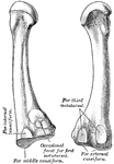
The second metatarsal of the left foot.

Metatarsal

The second metatarsal of the left foot.

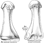
The third metatarsal of the left foot.

Metatarsal

The third metatarsal of the left foot.

The fourth metatarsal of the left foot.

Metatarsal

The fourth metatarsal of the left foot.

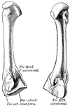
The fifth metatarsal of the left foot.

Metatarsal

The fifth metatarsal of the left foot.

The partial skeleton of a moa, an enormous flightless bird once native to New Zeland, now extinct.

Moa skeleton

The partial skeleton of a moa, an enormous flightless bird once native to New Zeland, now extinct.

Inferior turbinated bone, convex surface. The inferior turbinated bones are situated on the outer wall of each side of the nostril. Teach consists of a layer of thin, spongy bone, curled upon itself like a scroll.

Human Nostril Bone

Inferior turbinated bone, convex surface. The inferior turbinated bones are situated on the outer wall…
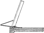
"It is worthy of note that, owing to the oblique direction in which the muscles are commonly inserted into the bones, much of their force is lost so far as producing movement is concerned. Suppose the log of wood in the diagram to be raised by pulling on the rope in the direction a; it is clear at first that the rope will act at a great disadvantage; most of the pull transmitted by it will be exerted against the pivot on which the log hinges, and only a small fraction be available for elevating the latter. But the more the log is lifted, as for example into the position indicated by the dotted lines, the more useful will be the direction of the pull, and the more of it will be spend on the log and the less lost unavailingly in merely increasing the pressure at the hinge." &mdash;Martin, 1917

Oblique Pull

"It is worthy of note that, owing to the oblique direction in which the muscles are commonly inserted…
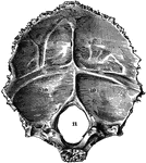
Occipital bone of the human skull, inner surface. It is situated at the back and base of the skull. Labels: 9,9, and 10,10, depression for reception of lobes of brain; 11, foramen magnum.

Occipital Bone of the Human Skull

Occipital bone of the human skull, inner surface. It is situated at the back and base of the skull.…
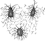
Oshyiodes of an adult fowl.

Oshyiodes of Adult Fowl

Oshyiodes of an adult fowl.

Nucleated none cells (osteoblasts) and their processes, contained in the one lacunae and their canaliculi respectively. From a section through the vertebra of an adult mouse.

Osteoblasts

Nucleated none cells (osteoblasts) and their processes, contained in the one lacunae and their canaliculi…
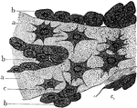
Osteoblasts from the parietal bone of a human embryo thirteen weeks old. Labels: a, bony septa with the cells of he lacunae; b, layers of osteoblasts; c, the latter in transition to bone corpuscles.

Osteoblasts from Embryo

Osteoblasts from the parietal bone of a human embryo thirteen weeks old. Labels: a, bony septa with…
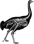
"Shows the powerful legs, small feet, and rudimentary wings of the bird; the obliquity at which the bones of the legs and wings are placed, and the comparatively small angles which two bones make at their point of junction."&mdash;Pettigrew, 1857

Skeleton of Ostrich

"Shows the powerful legs, small feet, and rudimentary wings of the bird; the obliquity at which the…
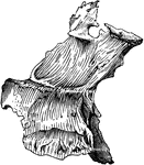
Palate bone. Palate bones form the back part of the roof of the mouth; part of the floor and outer wall of the nasal fossae, and a very small portion of the floor of the orbit.

Human Palate Bone

Palate bone. Palate bones form the back part of the roof of the mouth; part of the floor and outer wall…
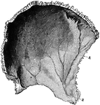
Parietal bone of the human skull, inner surface. The parietal bones form the greater part of the sides and roof of the skull. Labels: A, parietal depression; E, furrow for ramification of arteries.

Parietal Bone of the Human Skull

Parietal bone of the human skull, inner surface. The parietal bones form the greater part of the sides…
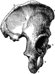
The Os Innominatum, or nameless bone, so called from bearing no resemblance to any known object, is a large irregular shaped bone, which, with its fellow of opposite side, forms the sides and front wall of the pelvic cavity. Labels: R, O, crest of ilium, just below O is seen the anterior superior spinous process; J, tuberosity of ischium; t, part of pubes, between J and T is seen the thyroid foramen: H, acetabulum, below H is seen end of ubic bone which, with its fellow of opposite side, forms the symphysis pubis.

Part of the Human Pelvic Bone

The Os Innominatum, or nameless bone, so called from bearing no resemblance to any known object, is…

Male pelvis (top) and female pelvis (bottom). The pelvis is stronger and more massively constructed than either the cranial or thoracic cavity. It is composed of 4 bones, the ossa innominata, forming sides and front, and the sacrum and coccyx, completing it behind. The female pelvis differs from that of the male in making it better adapted for giving birth. The most notable differences are that the female pelvis is wider in every direction, giving more room for the child to pass and shallower, which lessens the distance through which the child has to be propelled; and lastly, the bones are thinner and smoother.

Human Pelvis, Male and Female

Male pelvis (top) and female pelvis (bottom). The pelvis is stronger and more massively constructed…

"The bones of fishes are of a less dense and compact nature than in the higher order of animals; in some, indeed, they are wholly cartilaginous. The skeleton may in general be divided into four chief parts - the Vertical Column, the Head, the Repiratory Apparatus, and the Limbs" &mdash; Goodrich, 1859

Perch skeleton

"The bones of fishes are of a less dense and compact nature than in the higher order of animals; in…

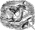
"Skeleton of the Perch's Skull." &mdash; Encyclopedia Britannica, 1893

Perch Skull

"Skeleton of the Perch's Skull." — Encyclopedia Britannica, 1893

"Lower view of skull of perch." &mdash; Encyclopedia Britannica, 1893

Perch Skull

"Lower view of skull of perch." — Encyclopedia Britannica, 1893

"Bones of the fore foot of existing Perissodactyle. Tapir (Tapirus indicus)." &mdash;The Encyclopedia Britannica, 1903

Perissodactyle

"Bones of the fore foot of existing Perissodactyle. Tapir (Tapirus indicus)." —The Encyclopedia…

"Bones of the fore foot of existing Perissodactyle. Rhinoceros (Rhinoceros Sumairensis)." &mdash;The Encyclopedia Britannica, 1903

Perissodactyle

"Bones of the fore foot of existing Perissodactyle. Rhinoceros (Rhinoceros Sumairensis)." —The…

"Pectoral arch and fore limb of the pike (Esox lucius), an osseous fish, showing scapulocoracoid, composed of Scp, scapula or hypercoracoid; c, posterior end of the outer margin of the scapulocoracoid; b, b, 1, 2, 3, 4, 5, five fin-rays or radialia; a, actinosts or basalia." -Whitney, 1911

Pike Scapulocoracoid

"Pectoral arch and fore limb of the pike (Esox lucius), an osseous fish, showing scapulocoracoid, composed…

The radial bone in the human arm.

Radius

The radial bone in the human arm.

"When a bone is broken, blood trickles out between the injured parts, and afterwards gives place to a sticky, watery fluid, which gradually becomes thicker, like a syrup or jelly. This is slowly replaced by a new bone structure and forms a kind of cement to gold together the broken ends." — Blaisedell, 1904

Broken Radius

"When a bone is broken, blood trickles out between the injured parts, and afterwards gives place to…
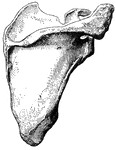
"The <em>shoulder-blade</em> is a large, flat, three-sided bone, which is placed on the upper and back part of the chest. On the outer side it has a saucerlike cavity on which the rounded head of the arm bone rests." &mdash; Blaisedell, 1904

Scapula

"The shoulder-blade is a large, flat, three-sided bone, which is placed on the upper and back…

The human scapula bone (shoulder blade). Labels: 1, glenoid cavity; 2, end of the spine of scapula.

The Human Scapula

The human scapula bone (shoulder blade). Labels: 1, glenoid cavity; 2, end of the spine of scapula.

This illustration shows the leg of a seal.
P. Pelvis, FE. Femur, TI. Tibia, FI. Fibula, TA. Tarsus, MT. Metatarsus, PH. Phalanges, OC. Os Calcis

Leg of Seal

This illustration shows the leg of a seal. P. Pelvis, FE. Femur, TI. Tibia, FI. Fibula, TA. Tarsus,…

This illustration shows a section of a human kidney (A, Cortical substance; B, Pyramids; C, Hilum; D, Pelvis; E, Ureter; F, Suprarenal capsule).
Kidneys are excretory organs whose function is to get rid of nitrogenous waste.

Section of Human Kidney

This illustration shows a section of a human kidney (A, Cortical substance; B, Pyramids; C, Hilum; D,…
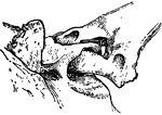
"A joint involving the principle of the shackle. Specifically, in anatomy, a kind of articulation found in the exoskeleton of some fishes, formed by the passing of a bony ring of one part through a perforation of another part, the two being thus movably linked together." &mdash;Whitney, 1889
<p>This illustration shows the shackle joint of a large spine with a bony plate of the skin of a siluroid fish.

Shackle Joint from the Exoskeleton of a Siluroid Fish

"A joint involving the principle of the shackle. Specifically, in anatomy, a kind of articulation found…