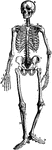
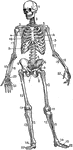
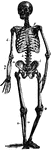
Clipart tagged: ‘bones’
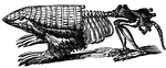
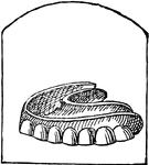
Armadillo - Endoskeleton and Exoskeleton or Dermoskeleton
An illustration of a pichiciago, a small burrowing armadillo. The front half of the animal is covered…
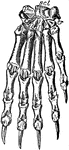
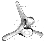
Bear Foot
"Palmar aspect of left fore foot of a black bear (Ursus americanus). scl, scapholunar; c, cuneiform;…
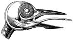
Curlew Skull
"Schizorhinal skull of curlew (top view), showing the long cleft, a, between upper and lower forks of…
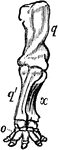
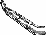
Anterior Extremity of Elephant
"Shows how the bones of the arm (q), forearm (q'x), and foot (o), are twisted to form an osseous screw."—Pettigrew,…
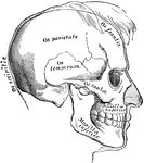
Eye Muscles
"The external bones of the temple are supposed to be removed in order to render visible the muscular…
Femur
Section of the femur. 1: External view; 2: Cellular portion at end; 3: Hollow in middle; 4: Thick shell…
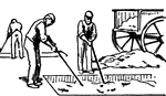
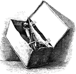
First Stage in Glue Manufacturing
This illustration shows the first stage in the manufacturing of glue. The material used for making the…
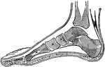
Side View of Bones in Foot
"The Foot is that part of the lower extremity below the leg on which we stand and walk. It is composed…
Side View of Bones in Foot
"The Foot is that part of the lower extremity below the leg on which we stand and walk. It is composed…
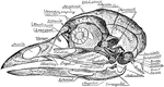
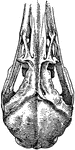
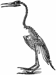
Skull of a Common Fowl
"Fig. 62 Skull of common fowl, enlarged. from nature by Dr. R.W. Shufeldt, U.S.A. The names of bones…
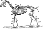
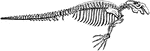
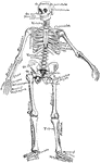
Horse Skeleton
"Skeleton of Horse (Equus caballus). fr, frontal bone; C, cervical vertebrae; D, dorsal vertebrae; L,…
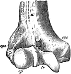
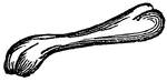
Humerus
"Anterior View, Distal End, of Right Humerus of a Man. H, humerus; epc, epicondyle, or external supracondyloid…
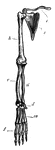
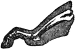
Chimp Limb
The forelimb of chimpanzee. (c) collar bone; (s) shoulder blade; (h) humerus; (r) radius; (u) ulna;…
Chimp Limb
The hindlimb of chimpanzee. (i) innominate bone; (f) thigh bone; (t) tibia; (s) fibula; (r) bones of…
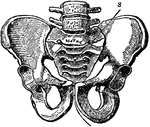
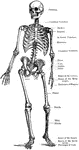
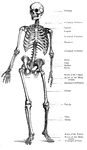
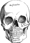
Pelvic Bone, Male
A bony structure located at the bottom of the spine. The human sacrum forms the back part of the pelvis,…
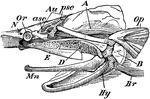
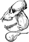
The Skull of a Paddle-Fish with the Beak Removed
"Skull of Spatularia, with the long beak removed, the anterior (asc) and posterior (psc) semicircular…
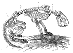
Beaver Skeleton
The skeleton of a beaver.(c) cervical region of vertebral column; (d) dorsal region; (b) lumbar region;…
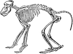
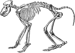
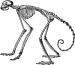
Chacma Baboon Skeleton
"The skeleton, more especially in the higher forms, is in the main similar to that of man, so that only…
Front View of Gorilla Skeleton
"The greatest absolute length of the fore—limb occurs in the gorilla and the orangutan. The humerus…
Side View of Skeleton of South American Spider Monkey
"In the other forms the number (vertebrae) varies between twenty and thirty three, the latter being…
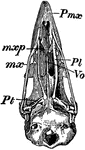
Bird Skull
"Schizognathous skull of common fowl. pmx, premaxilla; mxp, maxillopalatine; mx, maxilla; pl, palatine;…
Side View of Howler Monkey
A side view of the howler monkey skull. The monkey have four sharp canines, long teeth on skull, on…

Adult Male Orangutan Skull Viewed from Side
An illustration of an adult male orangutan viewed from the side. The orbit part of the skull is more…
Teeth
The series of small bones attached to the jaws of animals, or human beings, which serve the purpose…
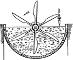
Wash Box for Removing Lime
This illustration shows a wash box used for removing lime from materials used in glue manufacturing.