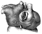
<em>L.A.</em>, left auricle; <em>R.A.</em>, right auricle; <em>A</em>, opening of the inferior vena cava; <em>B</em>, superior vena cava; <em>C</em>, cardiac vein of right auricle laid open; <em>D</em>, left pulmonary veins; <em>E</em>, eight pulmonary veins; <em>F</em>, muscular fibers surrounding the openings between the left auricle and left ventricle, and the right auricle and right ventricle; <em>G, H, K,</em> and <em>L</em>, muscular fibers surrounding the great veins of the heart." &mdash; Blaisedell, 1904

Muscular fibers of the auricle

L.A., left auricle; R.A., right auricle; A, opening of the inferior vena…

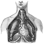
"<em>A</em>, a muscle which aids in pushing the food down the esophagus; <em>B</em>, esophagus; <em>C</em>, section of the right bronchus; <em>D</em>, two right pulmonary veins; <em>E</em>, great azygos vein crossing the esophagus to empty into the superior vena cava, <em>F</em>, thoracic duct; <em>H</em>, thoracic aorta; <em>K</em>, lower portion of the esophagus passing through the diaphragm; <em>L</em>, diaphragm as it appears in sectional view, enveloping the heart; <em>M</em>, inferior vena cava, passing through the diaphragm and emptying into the right auricle; <em>N</em>, right auricle; <em>O</em>, section of the right branch of the pulminary artery; <em>P</em>, aorta; <em>R</em>, superior vena cava; <em>S</em>, trachea." &mdash; Blaisedell, 1904

Lateral section of the chest

"A, a muscle which aids in pushing the food down the esophagus; B, esophagus; C,…

"<em>R.A.</em>, right auricle; <em>L.A.</em>, left auricle; <em>R.V.</em>, right ventricle; <em>L.V.</em>, left ventricle; <em>P.A.</i., pulmonary artery; <em>A</em>, pulmonary artery and vein of right lung; <em>B</em>, pulmonary artery and vein of left lung; <em>C</em>, carotid artery to head, showing branch of left subclavian artery; <em>D</em>, portal vein; <em>F</em>, hepatic artery; <em>G</em>, superior vena cava, bringing blood from head and upper limbs to right auricle." &mdash; Blaisedell, 1904

Diagram of the circulation of the blood

"R.A., right auricle; L.A., left auricle; R.V., right ventricle; L.V.,…
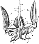
"Central organs of the circulaion, gills, and renal organs of Sepia officinalis. a, aorta; v, vena cava; v' visceral veins; c, systemic heart; d, dilatations of branchial veins on entering the heart; e, branchial hearts; b, branchiae; r, renal organs." &mdash; Encyclopedia Britannica, 1893

Cuttlefish Organs

"Central organs of the circulaion, gills, and renal organs of Sepia officinalis. a, aorta; v, vena cava;…
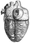
"<em>L.A.</em>, left auricle; <em>R.A.</em>, right auricle; <em>R.V.</em>, right ventricle; <em>A</em>, opening of the inferior vena cava; <em>B</em>, superior vena cava; <em>C</em>, right pulmonary veins; <em>D</em>, left pulmonary veins; <em>E</em>aorta; <em>F</em>, left branch of pulmonary artery; <em>G</em>, great cardiac vein; <em>H</em>middle cardiac vein. the cardiac lymphatics which follow the course of the cardiac veins are also shown." — Blaisedell, 1904

Posterior View of the Heart

"L.A., left auricle; R.A., right auricle; R.V., right ventricle; A,…

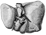
"The Liver seen from Below and Behind. <em>A</em> and </em>B</em>, smaller lobes of the liver; <em>C</em>, portal vein; <em>D</em> hepatic artery; <em>E</em>, inferior vena cava; <em>F</em>, trunk of the left hepatic vein; <em>G</em>, hepatic vein." &mdash; Blaisedell, 1904

Liver

"The Liver seen from Below and Behind. A and B, smaller lobes of the liver; C,…

"<em>L.A.</em>, left ventricle; <em>B</em>, right ventricle; <em>F</em>, pulmonary artery; <em>G</em>, aorta; <em>H</em>, arch of the aorta; <em>K</em>, innominate artery; <em>L</em>, right common carotid artery; <em>M</em>, right subclavian artery; <em>N</em>, thyroid cartilage forming upper portions of the larynx; <em>O</em>, trachea." &mdash; Blaisedell, 1904

Lungs

"L.A., left ventricle; B, right ventricle; F, pulmonary artery; G,…

"<em>A</em>, inferior vena cava, cut off just above the hepatic veins; <em>B</em>, superior vena cava, cut off just below the junction of the innominate veins; <em>C</em>, right and left innominate veins; <em>D</em>, left internal jugular vein. The veins, known as <em>azygos</em>, with some of their main branches, are shown. These veins connect the superior vena cava and the inferior vena cava." &mdash; Blaisedell, 1904

Vena cava

"A, inferior vena cava, cut off just above the hepatic veins; B, superior vena cava,…