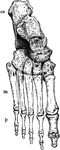
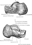
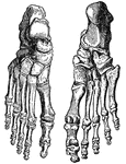
Bones of the Ankle and Foot
Bones of the Ankle and Foot. Labels: m, metatarsal bones; p, phalanges; ca, os calcis, or heel bone.
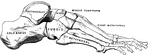
Ankle Joint and Foot
Vertical section of the ankle joint and foot. Labels: 1, tibia; 2, astragalus; 3, os calci; 4, scaphoides;…
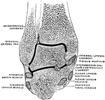
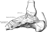
Coronal Section Through the Ankle Joint
Coronal section through the ankle joint and the calcaneo-astragaloid articulation.
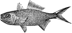
Anomalops Palpebratus
"A genus of fishes, typical of the family Anomalopidæ: so called from the remarkable structure…
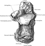
Artiodactyles
"Bones of fore foot of existing Artiodactyle. Pig (Sus scrofa)." —The Encyclopedia Britannica,…
Artiodactyles
"Bones of fore foot of existing Artiodactyle. Red Deer (Cervus elaphus)." —The Encyclopedia Britannica,…
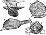
Forms of Bivalves
"Several Forms of Bivalves. A, Avicula; B, Pectunculus, with extended foot (a); C, Venus, with respiratory…
Circulation of blood
"Showing how the circulation of blood in the web of a frog's foot looks as seen under the microscope."…
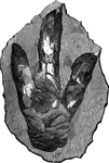
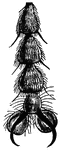
Footprint of brontozoum giganteum
A footprint of brontozoum giganeum, a now extinct relative of the cassowary. This example is eighteen…
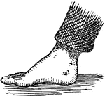
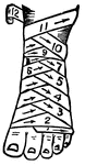
Club Foot
"A distortion or twisting of the foot by one or more of its muscles being permanently shortened. It…
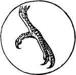
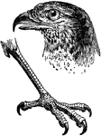
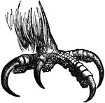
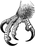
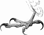
Foot of a Cuckoo
The foot of a Cuckoo, a bird belonging to the Scansores order. Scansores is an order of birds, popularly…
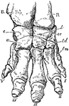
Elephant Foot
"Right fore foot of Indian Elephant. U, ulna; R, radius; c, cunelform; l, lunar; sc, scaphold; u, unciform;…
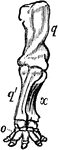
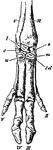
Anterior Extremity of Elephant
"Shows how the bones of the arm (q), forearm (q'x), and foot (o), are twisted to form an osseous screw."—Pettigrew,…

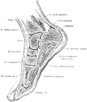
Frontal Section of Foot and Ankle
Frontal section of the right ankle and foot. Viewed from in front.
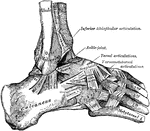
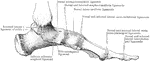
Ligaments of the Foot and Ankle
"The bones are fastened together, kept in place, and their movements limited, by tough and strong bands,…
Sagittal Section of Foot and Ankle
Sagittal section of the foot and ankle passing through the great toe.
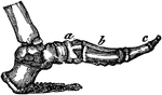
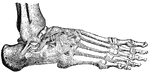
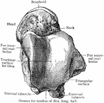
Side View of the Bone of the Foot
Bones of the foot, side view. In this figure the bones of the tarsus extend from the heel to a;…
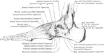
Outer Aspect of Foot Ligaments
Ligaments on outer aspect of ankle and on dorsum and outer aspects of foot.
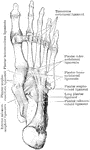

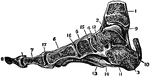
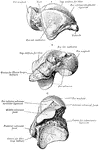
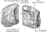
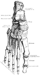
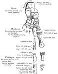
Bones of the Foot
The bones of the foot. Labels: Ca, Calcaneum, or heel bone; Ta, articular surface for tibia on the astragalus;…

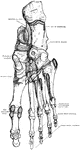
Bones of the Foot
Bones of the foot. At e d f g h are the 7 bones of the tarsus; at a are the 5 bones…