This illustration shows the plantigrade leg of a bear. Plantigrade means that the animal walks flat on its foot.
P. Pelvis, FE. Femur, TI. Tibia, FI. Fibula, TA. Tarsus, MT. Metatarsus, PH. Phalanges, OC. Os Calcis

Leg of Bear

This illustration shows the plantigrade leg of a bear. Plantigrade means that the animal walks flat…

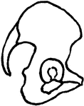
The chest and pelvis. 1: The hip-bones.

Chest and Pelvis

The chest and pelvis. 1: The hip-bones.

View of the diaphragm; 1, cavity of the thorax; 2, diaphragm separating the cavity of the thorax from that of the abdomen; 3, cavity of the pelvis.

Diaphragm

View of the diaphragm; 1, cavity of the thorax; 2, diaphragm separating the cavity of the thorax from…
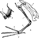
"A. Pelvis and bones of the leg of the Leon or Diver; i, Innominate bone; f, Thighbone (femur); r, Tibia; r, Fibula, together forming the shank; m, Tarso-metatarsus; p Phalanges of the toes. B, Tail of the Golden Eagle; s, Ploughshare-shaped bone, carrying the great tail-feathers." — Winston's Encyclopedia, 1919

Diver Bones

"A. Pelvis and bones of the leg of the Leon or Diver; i, Innominate bone; f, Thighbone (femur); r, Tibia;…
"Skeleton of Dugong: In which the pelvis is a mere bony ring." — Chambers, 1881

Dugong Skeleton

"Skeleton of Dugong: In which the pelvis is a mere bony ring." — Chambers, 1881

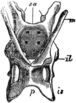
"The pelvis of the Echidna; sa, sacrum; il, illum; is, ischium; p, pubis; m, marsupial bone." — Chambers, 1881

Echidna Pelvis

"The pelvis of the Echidna; sa, sacrum; il, illum; is, ischium; p, pubis; m, marsupial bone." —…

Sagittal section in the median line of the female pelvis.

female Pelvis

Sagittal section in the median line of the female pelvis.

The pelvis and caudal vertebrae of an adult fowl.

Fowl Pelvis

The pelvis and caudal vertebrae of an adult fowl.

"Pelvis of a young grouse, showing three distinct bones. Il,P, ilium, ischium, pubis. In front of former a dorsal vertebra protrudes." Elliot Coues, 1884

The Pelvis of a Young Grouse

"Pelvis of a young grouse, showing three distinct bones. Il,P, ilium, ischium, pubis. In front of former…

"Pelvis of a heron (ardea herodias), nat. size, viewed from below; from nature by Dr. R.W. Shufeldt, U.S.A.   dl, dorso-lumbar vertebrae to and including the last one, sc; below sc, for the extent of the large black spaces (opposite the arrow) are the true sacral vertebrae; us, urosacral vertebrae (opposite the five oval black spaces; Il, ilium; Is, ischium; P, pubis; ob, obturator foramen. The arrow flies into the acetabulum.

The Pelvis of a Heron

"Pelvis of a heron (ardea herodias), nat. size, viewed from below; from nature by Dr. R.W. Shufeldt,…
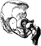
Line of pressure between hip and knee.

Line of Pressure Between Hip and Knee

Line of pressure between hip and knee.

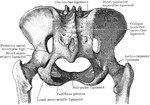
Ligaments and bones of the hip joint and pelvis. Labels: 1, posterior sacro-iliac ligament; 2, greater sacro sciatic ligament; 3, lesser sacro-sciatic ligament; 4, greater sacro-sciatic notch; 5, lesser sacro-sciatic notch; 6, cotyloid ligament around the acetabulum; 7, ligamentum teres; 8, line of attachment of the capsular ligament of the hip-joint, posteriorly; 9, obturator ligament.

Bones and Ligaments of the Hip and Pelvis

Ligaments and bones of the hip joint and pelvis. Labels: 1, posterior sacro-iliac ligament; 2, greater…

This illustration shows a front view of a human leg.
P. Pelvis, FE. Femur, TI. Tibia, FI. Fibula, TA. Tarsus, MT. Metatarsus, PH. Phalanges, OC. Os Calcis.

Human Leg (Front View)

This illustration shows a front view of a human leg. P. Pelvis, FE. Femur, TI. Tibia, FI. Fibula, TA.…

This illustration shows a human leg (front view), and comparative diagrams showing modifications of the leg:
1. Human, 2. Ox, 3. Horse, 4. Bird, 5. Frog, 6. Crocodile, 7. Seal, 8. Dog, 9. Bear
P. Pelvis, FE. Femur, TI. Tibia, FI. Fibula, TA. Tarsus, MT. Metatarsus, PH. Phalanges, OC. Os Calcis.

Human Leg (Front View), and Comparative Diagrams showing Modifications of the Leg

This illustration shows a human leg (front view), and comparative diagrams showing modifications of…

This illustration shows a side view of a human leg.
P. Pelvis, FE. Femur, TI. Tibia, FI. Fibula, TA. Tarsus, MT. Metatarsus, PH. Phalanges, OC. Os Calcis.

Human Leg (Side View)

This illustration shows a side view of a human leg. P. Pelvis, FE. Femur, TI. Tibia, FI. Fibula, TA.…
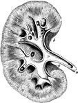
The ilum bone of the pelvis (hip region).

The Ilium

The ilum bone of the pelvis (hip region).

"<em>A</em>, pyramids of the kidney; <em>B</em>, apices, or papillae, of the pyramids; <em>C</em>, pelvis of the kidney; <em>D</em>, upper end of the ureter</em>." &mdash; Blaisedell, 1904

Kidney

"A, pyramids of the kidney; B, apices, or papillae, of the pyramids; C, pelvis…

Plan of a longitudinal section through the pelvis and substance of the right kidney. Labels: a, the cortical substance; b, broad part of the pyramids of Malpighi; c, the division of the pelvis named calyces, laid open; c', one of those unopened; d, summit of the pyramids of papillae projecting into calyces; p, pelvis or enlarged division of the ureter within the kidney; u, the ureter; s, the sinus; h, the hilus.

Kidney

Plan of a longitudinal section through the pelvis and substance of the right kidney. Labels: a, the…
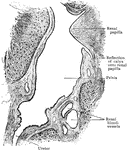
Sagittal section through sinus of child's kidney, showing lower part of pelvis and commencement of ureter.
Sagittal Section Through Sinus of Kidney

Sagittal section through sinus of child's kidney, showing lower part of pelvis and commencement of ureter.

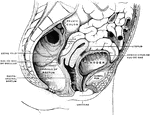
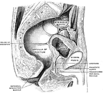
Sagittal section in the median line of the male pelvis.

Male Pelvis

Sagittal section in the median line of the male pelvis.

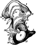
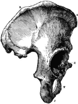
The Os Innominatum, or nameless bone, so called from bearing no resemblance to any known object, is a large irregular shaped bone, which, with its fellow of opposite side, forms the sides and front wall of the pelvic cavity. Labels: R, O, crest of ilium, just below O is seen the anterior superior spinous process; J, tuberosity of ischium; t, part of pubes, between J and T is seen the thyroid foramen: H, acetabulum, below H is seen end of ubic bone which, with its fellow of opposite side, forms the symphysis pubis.

Part of the Human Pelvic Bone

The Os Innominatum, or nameless bone, so called from bearing no resemblance to any known object, is…
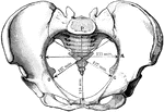
The diameters of the pelvic inlet in the female.

Pelvic Inlet

The diameters of the pelvic inlet in the female.

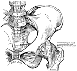
Pelvic joints as seen from behind.

Pelvic Joints Seen from Behind

Pelvic joints as seen from behind.

Pelvic joints as seen from the front.

Pelvic Joints Seen from Front

Pelvic joints as seen from the front.

The diameters of the pelvic outlet in the female.

Pelvic Outlet

The diameters of the pelvic outlet in the female.

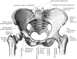
The anterior view of the articulations of the pelvis and hip.

Pelvis

The anterior view of the articulations of the pelvis and hip.

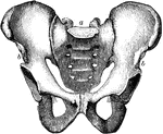
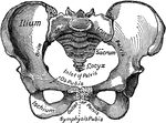
The pelvis of the human body. 1: Hip Bones; 2: Sacrum; 3: Coccyx.

Pelvis

The pelvis of the human body. 1: Hip Bones; 2: Sacrum; 3: Coccyx.

The Pelvis bone.

The Pelvis

The Pelvis bone.

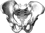
The pelvis.

Pelvis

The pelvis.

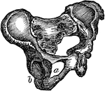
The pelvis. Labels: a, the sacrum; b, the right and the left innominatum.

The Pelvis

The pelvis. Labels: a, the sacrum; b, the right and the left innominatum.

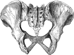
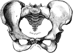
Female pelvis from before.

Female Pelvis from Before

Female pelvis from before.

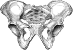
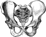
Male pelvis from before.

Male Pelvis from Before

Male pelvis from before.

The pelvis from behind.

Pelvis from Behind

The pelvis from behind.

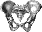
Female pelvis.

Female Pelvis

Female pelvis.

The female pelvis.

Female Pelvis

The female pelvis.

The female pelvis as seen from in front.

Female Pelvis

The female pelvis as seen from in front.

The male pelvis.

Male Pelvis

The male pelvis.

The male pelvis as seen from in front.

Male Pelvis

The male pelvis as seen from in front.

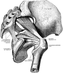
Muscles of the true pelvis on the right side, viewed from without and below. The quadratus having been removed, the gemelli are also not shown.

Muscles of Pelvis

Muscles of the true pelvis on the right side, viewed from without and below. The quadratus having been…
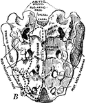
Side view of pelvis.

Side View of Pelvis

Side view of pelvis.

A large triangular bone as the base of the spine. Resides inbetween the two hip bones.

Bird Sacrum

A large triangular bone as the base of the spine. Resides inbetween the two hip bones.

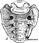
A large triangular bone as the base of the spine. Resides between the two hip bones.

Human Sacrum

A large triangular bone as the base of the spine. Resides between the two hip bones.

A large triangular bone as the base of the spine. Resides in between the two hip bones.

Human Sacrum

A large triangular bone as the base of the spine. Resides in between the two hip bones.