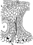
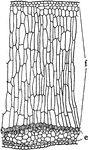
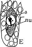
N. Odorata Epidermis
"C, portion of cross section of submerged stem of Nymphaea odorata, where there is no cutinized layer,…
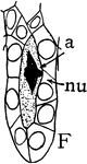
P. Japonica Epidermis
"A, cross section through upper half of leaf of Pyrus Japonica, showing cutinized layer of the outer…
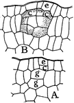
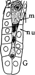
Plant Epidermis
"Two epidermal cells in cross section showing thickened outer wall differentiated into three layers,…
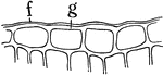
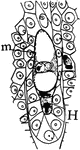
Russian Olive Leaf Epidermis
A cross section through the upper half of a Russian olive leaf, showing a "cutinized layer of the outer…
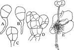
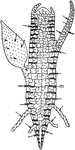
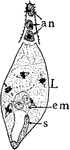
Fern Growth
"A, B, C, D, Successive stages of growth of prothallium from the spore in Osmunda cinnamomea; E, growth…
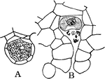
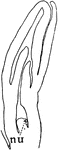
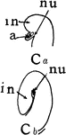
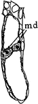
Fern Reproduction
"A, Antheridium containing sperm cells; B, archegonium containing an egg cell which has been found by…
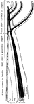
Indian Corn Food Circulation
"Diagram showing how, in Indian corn, the food from the upper and lower leaves finds its way into the…
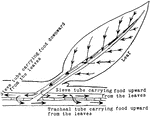
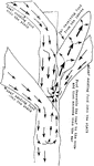
Leaf Food Circulation
"Diagram illustrating the descent of food from the leaf into the stem, and its circulation upward and…
Plant Food Digestion
"The digestion of the stored food and its ascent through the tracheal tubes when growth is resumed in…
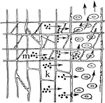
Plant Food Storage
"Diagram to show food from the leaves descending through the sieve tubes and being stored in the medullary…
Food Tissues
"Diagram to show the relation of the food-conducting tissues of the leaf to those of the stem; and in…
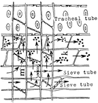
Sieve Tube Food Transportation
"Diagram showing the transport of food through the sieve tubes, medullary rays and tracheal tubes, and…
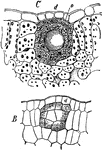
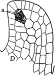
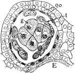
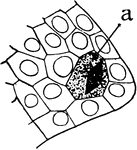
D. Fraxinella Gland Formation
"Formation of an interior, globular, lysigenous gland of the leaf of Dictamnus fraxinella. A, g, g and…
Leaf Gland
"Lysigenous gland in the leaf of Dictamnus fraxinella. B, young gland, with cells beginning to secrete…
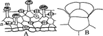
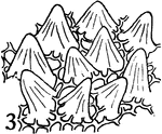
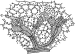
Leaf Glands
"Glands from Pinguicula. A, upper surface of leaf showing long-stalked gland at m, and short-stalked…
Leaf Glands
"Glands from the leaf of Ribes nigrum. A, young stage in the development of the gland where the cuticle…
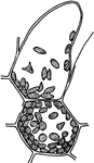
Orange Peel Glands
"Cross section through a portion of orange peel showing the cavity of an interior, globular gland at…
Guard Cell Experiment
"Diagram of apparatus showing how the guard cells draw apart; j, j, position of the rubber tubing when…
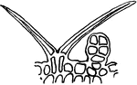
Hair, P. Zonale
"Glandular hair from the petiole of Pelargonium zonale. e, secretion from the globular gland on which…
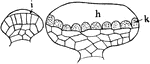
G. Pyriforme Hydatode
"One-celled hydatode of Gonocaryum pyriforme, seen in cross section at A, and from the surface in B."…
P. Sinensus Hydatode
"Radial longitudinal section through a hydatode from the leaf margin of Primula sinensis. i, upper,…
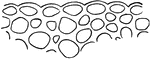
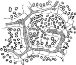
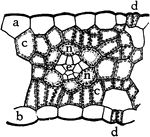
Intercellular Spaces of a Plant
"Diagram suggestive of the distribution of intercellular spaces throughout a plant. The heavy horizontal…
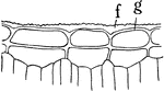
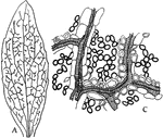
Intercellular Spaces of Leaf
"Showing intercellular spaces: f, between the palisade cells; e, in a leaf; g, border parenchyma; h,…
Leaf Architecture
"Diagram to show the architecture of a typical leaf in the region of one of the lateral veins. The shaded…
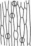
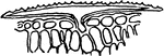
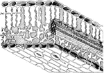
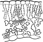
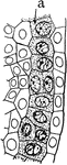
Indian Corn Leaf Cells
"Cross section of a portion of a leaf of Indian corn. a, upper and b, lower epidermis; c, c, palisade…
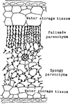
Rubber Leaf Cells
"Cross section through a portion of rubber leaf, showing the large percentage of water-storage tissues…
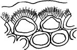
Leaf Epidermis
"Cross section of a portion of the blade of a leaf, showing upper epidermis at a, lower epidermis at…
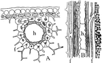
Leaf Resin Duct
"Resin duct in leaf of Pinus silvestris, in cross section at A, and in longitudinal section at B; h,…
Codonanthe Leaf Tissues
"Cross section of a portion of leaf of Codonanthe, showing the water-storage tissue at f, and the chlorophyll-bearing…
Cut Leaf Veins
"Showing the effect of cutting across the veins on the removal of food from the leaf. A, all of the…
Dicotyledon Leaf
A: "Camera-lucida drawing of a bleached leaf of a Dicotyledon, showing the course of the vascular bundles,…
E. Splendens Leaf
"Water-storage tracheids in the leaf of Euphorbia splendens. b, b, water-storage tracheids; d, mesophyll…
M. Forskalli Leaf
"Cross section of leaf of Mesembryanthemum Forskalii showing a large part of the leaf devoted to the…
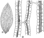
Monocotyledon Leaf
B: "Camera-lucida drawing of a bleached leaf of... a Monocotyledon, showing the anastomosis of the parallel…
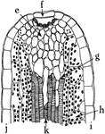
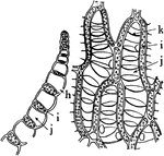
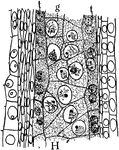
P. Commune Leaf
"Cross section through a portion of leaf of Polytrichum commune. b, chains of chlorophyll-bearing cells;…
Sphagnum Leaf
"Portion of leaf of Sphagnum, in cross section on the left, and surface view on the right. h, hole through…
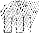
Plant Light Intake
"Diagram showing how the position of the chlorplasts against the vertical walls of the palisade cells…
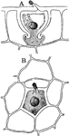
Megaspore Formation Stage 1
"Stages in the formation of the megaspore, its germination, fertilization of the egg and endosperm cells.…
Megaspore Formation Stage 10
"Stages in the formation of the megaspore, its germination, fertilization of the egg and endosperm cells.…
Megaspore Formation Stage 11
"Stages in the formation of the megaspore, its germination, fertilization of the egg and endosperm cells.…
Megaspore Formation Stage 12
"Stages in the formation of the megaspore, its germination, fertilization of the egg and endosperm cells.…
Megaspore Formation Stage 2
"Stages in the formation of the megaspore, its germination, fertilization of the egg and endosperm cells.…
Megaspore Formation Stage 3
"Stages in the formation of the megaspore, its germination, fertilization of the egg and endosperm cells.…
Megaspore Formation Stage 4
"Stages in the formation of the megaspore, its germination, fertilization of the egg and endosperm cells.…
Megaspore Formation Stage 5
"Stages in the formation of the megaspore, its germination, fertilization of the egg and endosperm cells.…
Megaspore Formation Stage 6
"Stages in the formation of the megaspore, its germination, fertilization of the egg and endosperm cells.…
Megaspore Formation Stage 7
"Stages in the formation of the megaspore, its germination, fertilization of the egg and endosperm cells.…
Megaspore Formation Stage 8
"Stages in the formation of the megaspore, its germination, fertilization of the egg and endosperm cells.…
Megaspore Formation Stage 9
"Stages in the formation of the megaspore, its germination, fertilization of the egg and endosperm cells.…
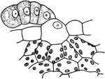
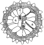
Microspore Anther
The anther and archesporium in the stages of "formation of anthers and pollen grains or microspores…
Microspore Anther Lobe
The cross section of a mature anther lobe in the stages of "formation of anthers and pollen grains or…
Microspore Anther Lobe
The cross section of a mature anther lobe in the stages of "formation of anthers and pollen grains or…
Microspore Archesporium
The archesporium in the stages of "formation of anthers and pollen grains or microspores of Silphium."…