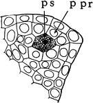
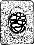
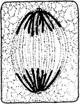
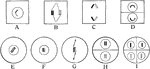
Microspore Cells
The primary sporogenous cell (ps) and the primary parietal layer (ppr) in the stages of "formation of…
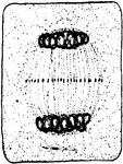
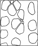
Microspore Cells
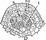
The sporogenous cells (s), tapetum (t), two parietal layers (oo) in the stages of "formation of anthers…
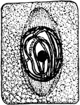
Microspore Cells
The sporogenous cells (s), tapetum (t), two parietal layers (oo) in the stages of "formation of anthers…
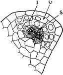
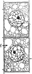
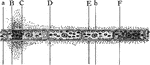
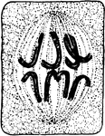
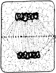
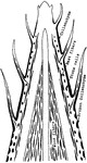
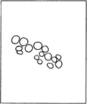
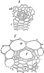
Microspore Layers
The inner layer (i), outer layer (o), sporogenous cells (s) in the stages of "formation of anthers and…
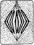
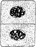
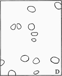
Microspore Pollen Grains
The pollen grains beginning to germinate in the stages of "formation of anthers and pollen grains or…
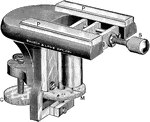
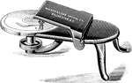
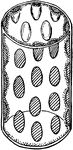
Microtome
"Simple microtome for clamping to table. P, P, plate glass ways for the section knife; S, milled-head…
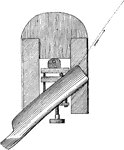
Microtome and Razor
"Showing the manner of holding the knife blade on the glass ways, and, by the arrow, the direction of…
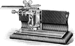
Sliding Microtome
"A sliding microtome of simple construction, adapted to cutting all kinds of sections." -Stevens, 1916
Mounting Plant Sections
"Turn table for cementing coverglasses to slides. For use where the mounting medium is glycerine or…
V. Faba Nucleus Division
"Nucleus dividing by simple constriction. From the lining of the embryo-sac of Vicia faba." -Stevens,…
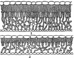
Oak Leaf Cross-Section
"Cross-sections of leaves of an oak (Quercus novimexicana), showing the effect of different light conditions…
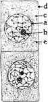
Onion Cells
"A, embryonic cells from onion root tip; d, plasmatic membrane; c, cytoplasm; a, nuclear membrane enclosing…
Onion Cells
"B, older (onion) cells farther back from the root tip. The cytoplasm is becoming vacuolate; f, vacuole."…
Palisade Cell
"Diagrammatic representation of a single palisade cell, with chloroplasts lining the walls." -Stevens,…
Palisade Cell
"Diagram to show the activities going on in a palisade cell. The arrows from the chloroplasts into the…

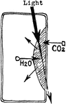
Palisade Cell Intake
"Diagram to show the intake of carbon dioxide by the palisade cells from the intercellular spaces, the…
Euphorbia Palisade Cells
"Starch grains from the palisade cells of a Euphorbia leaf." -Stevens, 1916
Photosynthesis
"Diagram to show the effect of different portions of the spectrum on photosynthesis. a to F, different…
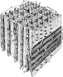
Magnified Pine
"Diagrammatic representation of a block of pine wood highly magnified. a, Early growth; b, late growth;…
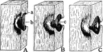
Pit Development
"Different stages in the development of a bordered pit. b, The original, thin, primary wall; a, the…
Plant Cell Division 1
First stage in plant cell division: Protophase 1; "Resting cell ready to begin division." -Stevens,…
Plant Cell Division 2
Second stage in plant cell division: Protophase 2; "the nuclear reticulum is assuming the form of a…
Plant Cell Division 3
Third stage in plant cell division: Protophase 3; "The nuclear thread has divided longitudinally throughout…
Plant Cell Division 4
Fourth stage in plant cell division: Protophase 4; "The nuclear membrane and the nucleolus have disappeared,…
Plant Cell Division 5
Fifth stage in plant cell division: Metaphase; "The metaphase, where the longitudinal halves of the…
Plant Cell Division 6
Sixth stage in plant cell division: Anaphase; "The anaphase, or movement of the chromosomes toward the…
Plant Cell Division 7
Seventh stage in plant cell division: Telophase; "Telophase where the chromosomes have begun to spin…
Plant Cell Division 8
Eighth stage in plant cell division: "The connecting fibers have spread out and come into contact with…
Plant Cell Division 9
Ninth and final stage in plant cell division: "A nuclear membrane has been formed about each daughter…
Plant Reproduction
"Showing method of association of paternal and maternal chromosomes, at A in all vegetative nuclei,…
Plant Tissues
"Diagram to show the topography and character of the tissues that evolved from the primary meristems.…
Plant Tissues
"Diagram showing additions to the primary tissues through the activity of the cambium and phellogen…
Plant Tissues
"Diagram showing the relation of this year's leaves to the wood of the current year." -Stevens, 1916
Plant Root Hair
"A single root hair on a large scale, showing that it is an outgrowth of an epidermal cell, and the…

Root Vessels
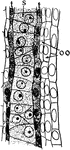
"Laticiferous vessels from the cortex of root of Scorozonora hispanica...B, smaller portion." -Stevens,…
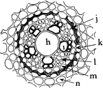
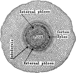
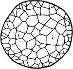
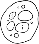
A. Ascalonicum Root
"Portion of a cross section of a root of Allium ascalonicum. h, large central, tracheal tube; i, xylem,…
Ryegrass Leaf with Sheath
Lolium perenne. A sheath, in botany is "the part of an expanded organ that is rolled around a stem or…
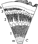
Plant Skeletal Tissues
"Diagram showing the progressive development of the skeletal tissues from the apex toward the base of…
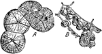
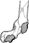
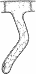
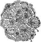
Sphaero-Crystals
"Sphaero-crystals of unilin from tuber of Dahlia variabilis. A, precipitated from an aqueous solution;…
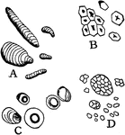
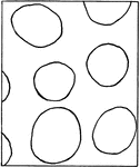
Starch
"Starch from different sources. A, curcuma starch; B, corn starch; C, tapioca starch; D, rice starch,…
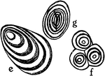
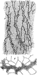
Starch Grain Striations
"Showing concentric and eccentric striations of starch grains. e, potato starch eccentrically striated;…
A. Capreolata Unusual Stem Growth
"Diagram showing some types of unusual growth in thickness. A, cross section through a four-year-old…
Bauhinia Unusual Stem Growth
"Diagram showing some types of unusual growth in thickness...B, cross section of stem of species of…
G. Scandens Unusual Stem Growth
"Diagram showing some types of unusual growth in thickness...C, portion of a cross section of stem of…
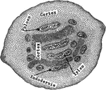
P. Galapageium Stem Tissue
The cross section of the stem of Pisdium Galapageium, which carries very little water.
Water Cress Stem Tissue
The cross section of the stem of water cress, which carries very little water.
Yellow Poplar Stem Tissue
The cross section of the stem of yellow poplar, which carries large amounts of water.
A. Pedaium Stem
"Adianium pedatum: transverse section of stem, showing the amphiphloic siphonostele." -Stevens, 1916
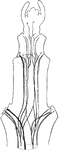
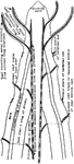
C. Viticella Stem
"Diagram showing the course of the vascular bundles in a stem of Clematis viticella. Median bundles…
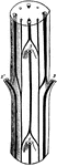
Cerastium Stem
"B, diagram of vascular bundles in external view and in cross section of the stem of Cerastium. The…
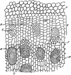
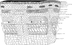
D. Marginata Stem
"Portion of a cross section throughout the stem of Dracaena marginata. P, parenchyma of cortex. V, meristematic…
G. Pubescens Stem
"Gleichenia pubescens: transverse section of stem, showing the protostele." -Stevens, 1916
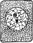
Ivy Stem
"Schizogenous resin duct in the young stem of ivy (Hedera helix), as seen in cross section. A, early,…
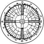
Juncus Stem
"A, cross section of stem of Juncus...the black lines traversing the section are really chains of cells…
P. Aquilina Stem
"Pteris aquilina: transverse section of stem, showing the polystele." -Stevens, 1916
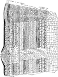
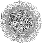
Palm Stem
"Cross section of a portion of palm stem. e, xylem; f, phloem portions of vascular bundle; g, sclerenchyma…