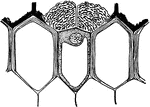
"Showing at f the felty body covering the passage way through the exodermis of the aerial root of Sobralia macrantha." -Stevens, 1916

S. Macrantha Aerial Root

"Showing at f the felty body covering the passage way through the exodermis of the aerial root of Sobralia…

"Portion of cross section through an aerial root of Stanhopea oculata. h, the velamen; i, exodermis; j, cortex; k, endodermis." -Stevens, 1916

S. Oculata Aerial Root

"Portion of cross section through an aerial root of Stanhopea oculata. h, the velamen; i, exodermis;…

"To show aleurone grains. A, cells from cotyledon of seed of garden bean; n, aleurone grains; m, starch; B, cells from endosperm of castor bean; a, aleurone grains; l, ground substance; k, crystalloid; j, globoid." -Stevens, 1916

Aleurone Grains

"To show aleurone grains. A, cells from cotyledon of seed of garden bean; n, aleurone grains; m, starch;…

"Diagram to show different plans in the distribution of bast fibers. A, bast a continuous cylinder in the pericycle; B, isolated strands of bast in the cortex and in the pericycle in front of each vascular bundle; C, a combination of A and B." -Stevens, 1916

Bast Fibers

"Diagram to show different plans in the distribution of bast fibers. A, bast a continuous cylinder in…

Developmental stages of bast fibers: "1 and 2, cross and longitudinal sections of primary meristem cells that are to become bast fibers." -Stevens, 1916

Bast Fibers 1 and 2

Developmental stages of bast fibers: "1 and 2, cross and longitudinal sections of primary meristem cells…

Developmental stages of bast fibers: 3 and 4, cross and longitudinal sections of meristem cells becoming bast fibers.

Bast Fibers 3 and 4

Developmental stages of bast fibers: 3 and 4, cross and longitudinal sections of meristem cells becoming…

Developmental stages of bast fibers: "5, longitudinal section of completed bast fibers. In 5 the stippling of the wall indicates lignification. Note that the walls have become pitted and the protoplasts have disappeared from the fibers." -Stevens, 1916

Bast Fibers 5

Developmental stages of bast fibers: "5, longitudinal section of completed bast fibers. In 5 the stippling…

"Camera-lucida outline of portion of cross section of cornstalk, showing at g bast fiber zone beneath the epidermis and surrounding the outermost vascular bundles." -Stevens, 1916

Cornstalk Bast Fibers

"Camera-lucida outline of portion of cross section of cornstalk, showing at g bast fiber zone beneath…
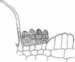
"Portion of a cross section through a nectariferous bract of Vicia sepium; n, nectar-secreting cells." -Stevens, 1916

V. Sepium Bract

"Portion of a cross section through a nectariferous bract of Vicia sepium; n, nectar-secreting cells."…

First developmental stage of sieve tubes, companion cells, and phloem parenchyma. "A, a and b, two rows of procambial cells; in c and d, a has divided longitudinal and c is to become companion cells; d, a sieve tube, and b, phloem parenchyma." -Stevens, 1916

Plant Cell Development 1

First developmental stage of sieve tubes, companion cells, and phloem parenchyma. "A, a and b, two rows…

Second developmental stage of sieve tubes, companion cells, and phloem parenchyma. "B, c, companion cells, and d, a beginning sieve tube from c and d, respectively in A. The cross-walls in d are pitted; b, phloem parenchyma grown larger." -Stevens, 1916

Plant Cell Development 2

Second developmental stage of sieve tubes, companion cells, and phloem parenchyma. "B, c, companion…

Third developmental stage of sieve tubes, companion cells, and phloem parenchyma. "The pits in the cross-walls of the sieve tubes become perforations, and the nuclei gone from the cells composing the tube." -Stevens, 1916

Plant Cell Development 3

Third developmental stage of sieve tubes, companion cells, and phloem parenchyma. "The pits in the cross-walls…

"Cross section through a water-absorbing scale of Tillandsia usneoides; a, a, water-absorbing cells partially filled with water." -Stevens, 1916

T. Usneoides Cell

"Cross section through a water-absorbing scale of Tillandsia usneoides; a, a, water-absorbing cells…

"D, cell of Oedogonium, with numerous chloroplasts." -Stevens, 1916

Oedogonium Chloroplast

"D, cell of Oedogonium, with numerous chloroplasts." -Stevens, 1916

"A, Pleurococcus, with chloroplast at c." -Stevens, 1916

Pleurococcus Chloroplast

"A, Pleurococcus, with chloroplast at c." -Stevens, 1916

"B, cell of Spirogyra, with spiral chloroplast at c, nucleus at n, and pyrenoid at e." -Stevens, 1916
Spirogyra Chloroplast

"B, cell of Spirogyra, with spiral chloroplast at c, nucleus at n, and pyrenoid at e." -Stevens, 1916

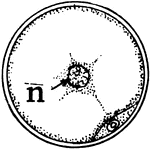
"C, cross section of Spirogyra cell, with nucleus at n, and section of chloroplast and pyrenoid below." -Stevens, 1916

Spirogyra Chloroplast

"C, cross section of Spirogyra cell, with nucleus at n, and section of chloroplast and pyrenoid below."…
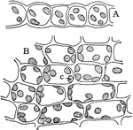
"Cross section, A, and surface view, B, of a leaf of common moss, showing chloroplasts, c." -Stevens, 1916

Moss Leaf Chloroplasts

"Cross section, A, and surface view, B, of a leaf of common moss, showing chloroplasts, c." -Stevens,…

"Enzyme-secreting cells of date cotyledon." -Stevens, 1916

Date Cotyledon

"Enzyme-secreting cells of date cotyledon." -Stevens, 1916

"Enzyme-secreting cells of...cotyledon of Indian corn." -Stevens, 1916

Indian Corn Cotyledon

"Enzyme-secreting cells of...cotyledon of Indian corn." -Stevens, 1916

"Different forms of crystals of calcium oxalate. A, from the petiole of Begonia manicata." -Stevens, 1916

Calcium Oxalate Crystals

"Different forms of crystals of calcium oxalate. A, from the petiole of Begonia manicata." -Stevens,…

"Cystoliths from the leaf of Ficus carica. A, complete cystolith; B, cystolith from which the calcium carbonate has been removed for use in other parts of the plant. B is from a leaf that had fallen off in autumn." -Stevens, 1916

Cystoliths

"Cystoliths from the leaf of Ficus carica. A, complete cystolith; B, cystolith from which the calcium…

"Longitudinal section through a digestive gland of Drosera rotundifolia." -Stevens, 1916

D. Rotundifolia Digestive Gland

"Longitudinal section through a digestive gland of Drosera rotundifolia." -Stevens, 1916
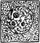
"Stages in the division of a grandmother cell of microspores or pollen grains of a lily, somewhat diagrammatic. 1, resting stage of a grandmother cell." -Stevens, 1916

Division of Grandmother Cell Stage 1

"Stages in the division of a grandmother cell of microspores or pollen grains of a lily, somewhat diagrammatic.…
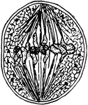
"Stages in the division of a grandmother cell of microspores or pollen grains of a lily, somewhat diagrammatic. 10, bipolar spindle with double chromosomes aligned at the equator." -Stevens, 1916

Division of Grandmother Cell Stage 10

"Stages in the division of a grandmother cell of microspores or pollen grains of a lily, somewhat diagrammatic.…
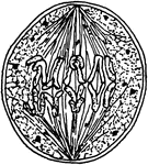
"Stages in the division of a grandmother cell of microspores or pollen grains of a lily, somewhat diagrammatic. 11, reduction division, the double chromosomes separating and at the same time showing longitudinal division, with the longitudinal halves widely apart at their equatorial ends." -Stevens, 1916

Division of Grandmother Cell Stage 11

"Stages in the division of a grandmother cell of microspores or pollen grains of a lily, somewhat diagrammatic.…
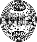
"Stages in the division of a grandmother cell of microspores or pollen grains of a lily, somewhat diagrammatic. 12, formation of daughter nuclei, which are really the nuclei of the mother cells of the spores." -Stevens, 1916

Division of Grandmother Cell Stage 12

"Stages in the division of a grandmother cell of microspores or pollen grains of a lily, somewhat diagrammatic.…
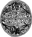
"Stages in the division of a grandmother cell of microspores or pollen grains of a lily, somewhat diagrammatic. 13, division of mother-cell nuclei, the longitudinally split chromosomes seen in 11 again appearing." -Stevens, 1916

Division of Grandmother Cell Stage 13

"Stages in the division of a grandmother cell of microspores or pollen grains of a lily, somewhat diagrammatic.…
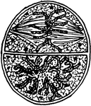
"Stages in the division of a grandmother cell of microspores or pollen grains of a lily, somewhat diagrammatic. 14, split chromosomes of 13 aligned at the equator ready for the longitudinal halves of each to be drawn apart to opposite poles." -Stevens, 1916

Division of Grandmother Cell Stage 14

"Stages in the division of a grandmother cell of microspores or pollen grains of a lily, somewhat diagrammatic.…
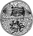
"Stages in the division of a grandmother cell of microspores or pollen grains of a lily, somewhat diagrammatic. 15, separation of the longitudinal halves." -Stevens, 1916

Division of Grandmother Cell Stage 15

"Stages in the division of a grandmother cell of microspores or pollen grains of a lily, somewhat diagrammatic.…
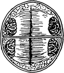
"Stages in the division of a grandmother cell of microspores or pollen grains of a lily, somewhat diagrammatic. 16, formation of granddaughter nuclei, or nuclei of the spores." -Stevens, 1916

Division of Grandmother Cell Stage 16

"Stages in the division of a grandmother cell of microspores or pollen grains of a lily, somewhat diagrammatic.…
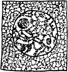
"Stages in the division of a grandmother cell of microspores or pollen grains of a lily, somewhat diagrammatic. 2, nuclear thread becoming arranged into parallel threads." -Stevens, 1916

Division of Grandmother Cell Stage 2

"Stages in the division of a grandmother cell of microspores or pollen grains of a lily, somewhat diagrammatic.…
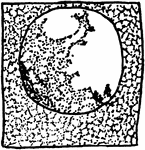
"Stages in the division of a grandmother cell of microspores or pollen grains of a lily, somewhat diagrammatic. 3, synaptic stage." -Stevens, 1916

Division of Grandmother Cell Stage 3

"Stages in the division of a grandmother cell of microspores or pollen grains of a lily, somewhat diagrammatic.…
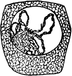
"Stages in the division of a grandmother cell of microspores or pollen grains of a lily, somewhat diagrammatic. 4, parallel threads uniting." -Stevens, 1916

Division of Grandmother Cell Stage 4

"Stages in the division of a grandmother cell of microspores or pollen grains of a lily, somewhat diagrammatic.…
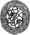
"Stages in the division of a grandmother cell of microspores or pollen grains of a lily, somewhat diagrammatic. 5, double threads so united as to appear as one." -Stevens, 1916

Division of Grandmother Cell Stage 5

"Stages in the division of a grandmother cell of microspores or pollen grains of a lily, somewhat diagrammatic.…
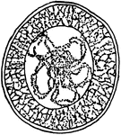
"Stages in the division of a grandmother cell of microspores or pollen grains of a lily, somewhat diagrammatic. 7, thread transversely segmented into double chromosomes." -Stevens, 1916

Division of Grandmother Cell Stage 7

"Stages in the division of a grandmother cell of microspores or pollen grains of a lily, somewhat diagrammatic.…
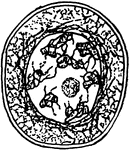
"Stages in the division of a grandmother cell of microspores or pollen grains of a lily, somewhat diagrammatic. 8, diakinesis, that is, chromosomes dispersed about the nuclear membrane." -Stevens, 1916

Division of Grandmother Cell Stage 8

"Stages in the division of a grandmother cell of microspores or pollen grains of a lily, somewhat diagrammatic.…
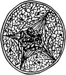
"Stages in the division of a grandmother cell of microspores or pollen grains of a lily, somewhat diagrammatic. 9, multipolar spindle stage." -Stevens, 1916

Division of Grandmother Cell Stage 9

"Stages in the division of a grandmother cell of microspores or pollen grains of a lily, somewhat diagrammatic.…

"Different forms of epidermal outgrowths. 1, hooked hair from Phaseolus multiflorus; 2, climbing hair from stem of Humulus Lupulus; 3, rod-like wax coating from the stem of Saccharum offininarum; 4, climbing hair of Lossa hispida; 5, stinging hair of Urtica urens." -Stevens, 1916

Epidermal Outgrowths

"Different forms of epidermal outgrowths. 1, hooked hair from Phaseolus multiflorus; 2, climbing hair…
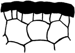
"B, cross section through skin of apple." -Stevens, 1916

Apple Epidermis

"B, cross section through skin of apple." -Stevens, 1916

"A, portion of cross section of leaf of Avicennia growing in salty soil; outer wall of epidermis very thick." -Stevens, 1916

Avicennia Epidermis

"A, portion of cross section of leaf of Avicennia growing in salty soil; outer wall of epidermis very…
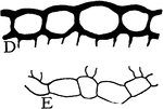
"D, upper, and E, lower epidermis of leaf of Hibiscus moscheutos." -Stevens, 1916

H. Moscheutos Epidermis

"D, upper, and E, lower epidermis of leaf of Hibiscus moscheutos." -Stevens, 1916

"C, cross section through upper half of petal of Japan Quince." -Stevens, 1916

Japan Quince Epidermis

"C, cross section through upper half of petal of Japan Quince." -Stevens, 1916

"Multiple epidermis of leaf of mangrove in cross section. This serves as a water reservoir, and the relatively thick walls of the inner cells reenforce the protective power of the outer layer." -Stevens, 1916

Multiple Epidermis

"Multiple epidermis of leaf of mangrove in cross section. This serves as a water reservoir, and the…

"C, portion of cross section of submerged stem of Nymphaea odorata, where there is no cutinized layer, and the cuticle is a hardly distinguishable film." -Stevens, 1916

N. Odorata Epidermis

"C, portion of cross section of submerged stem of Nymphaea odorata, where there is no cutinized layer,…

"A, cross section through upper half of leaf of Pyrus Japonica, showing cutinized layer of the outer wall at f, and cellulose layer at g." -Stevens, 1916

P. Japonica Epidermis

"A, cross section through upper half of leaf of Pyrus Japonica, showing cutinized layer of the outer…

A cross section through the upper half of a Russian olive leaf, showing a "cutinized layer of the outer wall at f, and cellulose layer at g." -Stevens, 1916

Russian Olive Leaf Epidermis

A cross section through the upper half of a Russian olive leaf, showing a "cutinized layer of the outer…
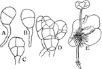
"A, B, C, D, Successive stages of growth of prothallium from the spore in Osmunda cinnamomea; E, growth of fern plant from fertilized egg within the prothallium of Osmunda Claytoniana." -Stevens, 1916

Fern Growth

"A, B, C, D, Successive stages of growth of prothallium from the spore in Osmunda cinnamomea; E, growth…
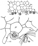
"Fern prothallus; cross-sections showing antheridia (an), sperms (sp), and rhizoids (rh). Below at the right is a sperm (sp) greatly enlarged." -Gager, 1916

Fern Prothallus

"Fern prothallus; cross-sections showing antheridia (an), sperms (sp), and rhizoids (rh). Below at the…
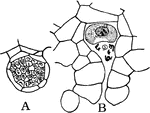
"A, Antheridium containing sperm cells; B, archegonium containing an egg cell which has been found by five sperm cells. All from Osmunda cinnamomea." -Stevens, 1916

Fern Reproduction

"A, Antheridium containing sperm cells; B, archegonium containing an egg cell which has been found by…

"Diagram showing how, in Indian corn, the food from the upper and lower leaves finds its way into the ears." -Stevens, 1916

Indian Corn Food Circulation

"Diagram showing how, in Indian corn, the food from the upper and lower leaves finds its way into the…
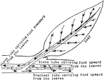
"Diagram illustrating the descent of food from the leaf into the stem, and its circulation upward and downward through the sieve tubes, and upward through the tracheal tissues." -Stevens, 1916

Leaf Food Circulation

"Diagram illustrating the descent of food from the leaf into the stem, and its circulation upward and…
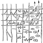
"The digestion of the stored food and its ascent through the tracheal tubes when growth is resumed in the spring." -Stevens, 1916

Plant Food Digestion

"The digestion of the stored food and its ascent through the tracheal tubes when growth is resumed in…
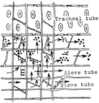
"Diagram to show food from the leaves descending through the sieve tubes and being stored in the medullary ray cells and xylem parenchyma." -Stevens, 1916

Plant Food Storage

"Diagram to show food from the leaves descending through the sieve tubes and being stored in the medullary…

"Diagram to show the relation of the food-conducting tissues of the leaf to those of the stem; and in the stem the relation of these tissues of one year to those of preceding years. The dotted line between phloem and xylem stands for the cambium. The figures at the bottom of the diagram indicate the age in years of the zones of tissues in phloem and xylem." -Stevens, 1916

Food Tissues

"Diagram to show the relation of the food-conducting tissues of the leaf to those of the stem; and in…

"Diagram showing the transport of food through the sieve tubes, medullary rays and tracheal tubes, and its storage in the parenchyma cells of the wood and bark. The black bodies in the cells indicate stored food." -Stevens, 1916

Sieve Tube Food Transportation

"Diagram showing the transport of food through the sieve tubes, medullary rays and tracheal tubes, and…
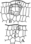
"Formation of an interior, globular, lysigenous gland of the leaf of Dictamnus fraxinella. A, g, g and c, mother cells of the gland; c, from the protoderm, and g, g, from the fundamental tissue. B, older stage where the cells have begun to form the secretion." -Stevens, 1916

D. Fraxinella Gland Formation

"Formation of an interior, globular, lysigenous gland of the leaf of Dictamnus fraxinella. A, g, g and…
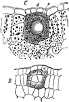
"Lysigenous gland in the leaf of Dictamnus fraxinella. B, young gland, with cells beginning to secrete oil; C, mature gland where the secreting cells have broken down and left their secretion within the cavity thus formed; o, large drop of secreted oil." -Stevens, 1916

Leaf Gland

"Lysigenous gland in the leaf of Dictamnus fraxinella. B, young gland, with cells beginning to secrete…

"Glands from Pinguicula. A, upper surface of leaf showing long-stalked gland at m, and short-stalked gland at n. B, cross section through a short-stalked gland." -Stevens, 1916

Leaf Glands

"Glands from Pinguicula. A, upper surface of leaf showing long-stalked gland at m, and short-stalked…

"Glands from the leaf of Ribes nigrum. A, young stage in the development of the gland where the cuticle is already being pushed up by the secretion, i. B, complete gland; k, secreting cells; h, cavity between the secreting cells and cuticle occupied by the secretion." -Stevens, 1916

Leaf Glands

"Glands from the leaf of Ribes nigrum. A, young stage in the development of the gland where the cuticle…