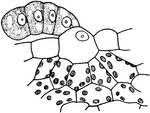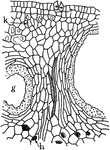
"Cross section through a portion of orange peel showing the cavity of an interior, globular gland at g; crystals of hesperidin at h; calcium oxalate crystals at k." -Stevens, 1916

Orange Peel Glands

"Cross section through a portion of orange peel showing the cavity of an interior, globular gland at…

"Diagram of apparatus showing how the guard cells draw apart; j, j, position of the rubber tubing when the water pressure is turned on." -Stevens, 1916

Guard Cell Experiment

"Diagram of apparatus showing how the guard cells draw apart; j, j, position of the rubber tubing when…

"Glandular hair from the petiole of Pelargonium zonale. e, secretion from the globular gland on which it rests." -Stevens, 1916

Hair, P. Zonale

"Glandular hair from the petiole of Pelargonium zonale. e, secretion from the globular gland on which…

"One-celled hydatode of Gonocaryum pyriforme, seen in cross section at A, and from the surface in B." -Stevens, 1916

G. Pyriforme Hydatode

"One-celled hydatode of Gonocaryum pyriforme, seen in cross section at A, and from the surface in B."…
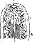
"Hydatode from the leaf of Phaseolus multiflorus." -Stevens, 1916

P. Multiflorus Hydatode

"Hydatode from the leaf of Phaseolus multiflorus." -Stevens, 1916

"Radial longitudinal section through a hydatode from the leaf margin of Primula sinensis. i, upper, and j, lower epidermis; h, palisade cell; e, thin-walled parenchyma, called epithem; g, intercellular space; f, guard cell of a water stoma; k, tracheal elements." -Stevens, 1916

P. Sinensus Hydatode

"Radial longitudinal section through a hydatode from the leaf margin of Primula sinensis. i, upper,…

"Diagram suggestive of the distribution of intercellular spaces throughout a plant. The heavy horizontal lines in stem and root indicate intercellular spaces in medullary rays. The arrows indicate the entrance of gasses through the stomata of the leaves and stomata and lenticels of the roots and everywhere over the younger roots where waterproofing is slight." -Stevens, 1916

Intercellular Spaces of a Plant

"Diagram suggestive of the distribution of intercellular spaces throughout a plant. The heavy horizontal…
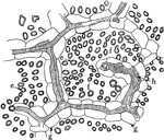
"Showing intercellular spaces: f, between the palisade cells; e, in a leaf; g, border parenchyma; h, tracheal elements of the vein." -Stevens, 1916

Intercellular Spaces of Leaf

"Showing intercellular spaces: f, between the palisade cells; e, in a leaf; g, border parenchyma; h,…
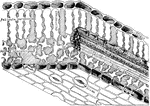
"Diagram to show the architecture of a typical leaf in the region of one of the lateral veins. The shaded parts amongst the palisade and spongy parenchyma represent intercellular spaces." -Stevens, 1916

Leaf Architecture

"Diagram to show the architecture of a typical leaf in the region of one of the lateral veins. The shaded…
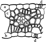
"Cross section of a portion of a leaf of Indian corn. a, upper and b, lower epidermis; c, c, palisade cells; n, border parenchyma containing starch within its chloroplasts; e, vascular bundle; d, d, stomata." -Stevens, 1916

Indian Corn Leaf Cells

"Cross section of a portion of a leaf of Indian corn. a, upper and b, lower epidermis; c, c, palisade…
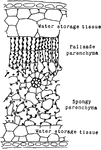
"Cross section through a portion of rubber leaf, showing the large percentage of water-storage tissues on both sides of the leaf, and the relation of the palisade and spongy parenchyma to the lateral veins." -Stevens, 1916

Rubber Leaf Cells

"Cross section through a portion of rubber leaf, showing the large percentage of water-storage tissues…
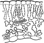
"Cross section of a portion of the blade of a leaf, showing upper epidermis at a, lower epidermis at b, palisade parenchyma at c, spongy parenchyma at d, and tracheids from the end of a vein at e." -Stevens, 1916

Leaf Epidermis

"Cross section of a portion of the blade of a leaf, showing upper epidermis at a, lower epidermis at…

"Resin duct in leaf of Pinus silvestris, in cross section at A, and in longitudinal section at B; h, cavity surrounded by the secreting cells; f, f, sclerenchyma fibers surrounding and protecting the duct." -Stevens, 1916

Leaf Resin Duct

"Resin duct in leaf of Pinus silvestris, in cross section at A, and in longitudinal section at B; h,…

"Cross section of a portion of leaf of Codonanthe, showing the water-storage tissue at f, and the chlorophyll-bearing tissues at e." -Stevens, 1916

Codonanthe Leaf Tissues

"Cross section of a portion of leaf of Codonanthe, showing the water-storage tissue at f, and the chlorophyll-bearing…

"Showing the effect of cutting across the veins on the removal of food from the leaf. A, all of the main veins are cut across near their bases; B, the mid-vein alone has been severed. The stippled areas indicated the starch reaction with the iodine test." -Stevens, 1916

Cut Leaf Veins

"Showing the effect of cutting across the veins on the removal of food from the leaf. A, all of the…
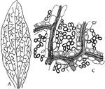
A: "Camera-lucida drawing of a bleached leaf of a Dicotyledon, showing the course of the vascular bundles, and how end free in the mesophyll." C:magnified detail. -Stevens, 1916

Dicotyledon Leaf

A: "Camera-lucida drawing of a bleached leaf of a Dicotyledon, showing the course of the vascular bundles,…
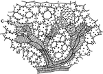
"Water-storage tracheids in the leaf of Euphorbia splendens. b, b, water-storage tracheids; d, mesophyll cells; c, branch from a milk tube." -Stevens, 1916

E. Splendens Leaf

"Water-storage tracheids in the leaf of Euphorbia splendens. b, b, water-storage tracheids; d, mesophyll…
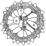
"Cross section of leaf of Mesembryanthemum Forskalii showing a large part of the leaf devoted to the storage of water. a, water-storage cells; b, chlorophyll-bearing cells; c, crystal of calcium oxalate." -Stevens, 1916

M. Forskalli Leaf

"Cross section of leaf of Mesembryanthemum Forskalii showing a large part of the leaf devoted to the…
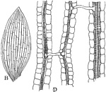
B: "Camera-lucida drawing of a bleached leaf of... a Monocotyledon, showing the anastomosis of the parallel veins by means of slender lateral branches." D: magnified detail. -Stevens, 1916

Monocotyledon Leaf

B: "Camera-lucida drawing of a bleached leaf of... a Monocotyledon, showing the anastomosis of the parallel…

"Cross section through a portion of leaf of Polytrichum commune. b, chains of chlorophyll-bearing cells; d, bast fibers." -Stevens, 1916

P. Commune Leaf

"Cross section through a portion of leaf of Polytrichum commune. b, chains of chlorophyll-bearing cells;…
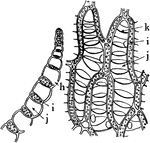
"Portion of leaf of Sphagnum, in cross section on the left, and surface view on the right. h, hole through the wall; i, chlorophyll-bearing cells; j, water-storage and water-conducting cells, with annular thickenings at k." -Stevens, 1916

Sphagnum Leaf

"Portion of leaf of Sphagnum, in cross section on the left, and surface view on the right. h, hole through…
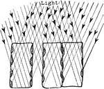
"Diagram showing how the position of the chlorplasts against the vertical walls of the palisade cells exposes them to good advantage to light from all quarters of the sky." -Stevens, 1916

Plant Light Intake

"Diagram showing how the position of the chlorplasts against the vertical walls of the palisade cells…

"Stages in the formation of the megaspore, its germination, fertilization of the egg and endosperm cells. A, beginning of ovule at o." -Stevens, 1916

Megaspore Formation Stage 1

"Stages in the formation of the megaspore, its germination, fertilization of the egg and endosperm cells.…

"Stages in the formation of the megaspore, its germination, fertilization of the egg and endosperm cells. J, continued nuclear division has resulted in eight, namely, three antipodal nuclei, an, at the upper end of the megaspore cavity, two polar nuclei, l, in process of fusion, egg cell, eg, two synergids, s, s." -Stevens, 1916

Megaspore Formation Stage 10

"Stages in the formation of the megaspore, its germination, fertilization of the egg and endosperm cells.…
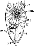
"Stages in the formation of the megaspore, its germination, fertilization of the egg and endosperm cells. K, showing only the lower half of the megaspore or embryo-sac cavity, with pollen tube, pt, and male nucleus, mn, fusing with egg cell, eg, and second male nucleus, mn2, uniting with primary endosperm nucleus, pe; s, synergid." -Stevens, 1916

Megaspore Formation Stage 11

"Stages in the formation of the megaspore, its germination, fertilization of the egg and endosperm cells.…

"Stages in the formation of the megaspore, its germination, fertilization of the egg and endosperm cells. L, later stage in the embryo-sac, an, antipodal cells, em, beginning of the embryo by division of fertilized egg cell; five dividing nuclei descended from primary endosperm nucleus." -Stevens, 1916

Megaspore Formation Stage 12

"Stages in the formation of the megaspore, its germination, fertilization of the egg and endosperm cells.…

"Stages in the formation of the megaspore, its germination, fertilization of the egg and endosperm cells. B, nu, nucellus" -Stevens, 1916

Megaspore Formation Stage 2

"Stages in the formation of the megaspore, its germination, fertilization of the egg and endosperm cells.…

"Stages in the formation of the megaspore, its germination, fertilization of the egg and endosperm cells. Ca, nu, nucellus, a, archesporium; in, forward growth of tissue to form the integument; Cb, nu, nucellus; in, integument." -Stevens, 1916

Megaspore Formation Stage 3

"Stages in the formation of the megaspore, its germination, fertilization of the egg and endosperm cells.…
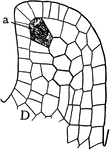
"Stages in the formation of the megaspore, its germination, fertilization of the egg and endosperm cells. D, more highly magnified drawing of o in A showing archesporial cell at a." -Stevens, 1916

Megaspore Formation Stage 4

"Stages in the formation of the megaspore, its germination, fertilization of the egg and endosperm cells.…
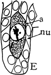
"Stages in the formation of the megaspore, its germination, fertilization of the egg and endosperm cells. E, a, archesporial cell enlarged and ready for division; nu, tissue of the nucellus." -Stevens, 1916

Megaspore Formation Stage 5

"Stages in the formation of the megaspore, its germination, fertilization of the egg and endosperm cells.…

"Stages in the formation of the megaspore, its germination, fertilization of the egg and endosperm cells. F, a, archesporial cell dividing, nu, nucellus." -Stevens, 1916

Megaspore Formation Stage 6

"Stages in the formation of the megaspore, its germination, fertilization of the egg and endosperm cells.…

"Stages in the formation of the megaspore, its germination, fertilization of the egg and endosperm cells. G, row of four megaspores descended from the archesporial cell, only the upper one, m, functional, the other soon disintegrate." -Stevens, 1916

Megaspore Formation Stage 7

"Stages in the formation of the megaspore, its germination, fertilization of the egg and endosperm cells.…

"Stages in the formation of the megaspore, its germination, fertilization of the egg and endosperm cells. H, megaspore, m, enlarging, the others disappearing." -Stevens, 1916

Megaspore Formation Stage 8

"Stages in the formation of the megaspore, its germination, fertilization of the egg and endosperm cells.…

"Stages in the formation of the megaspore, its germination, fertilization of the egg and endosperm cells. I, nucleus of the megaspore, divided into two, md." -Stevens, 1916

Megaspore Formation Stage 9

"Stages in the formation of the megaspore, its germination, fertilization of the egg and endosperm cells.…

The anther and archesporium in the stages of "formation of anthers and pollen grains or microspores of Silphium." -Stevens, 1916

Microspore Anther

The anther and archesporium in the stages of "formation of anthers and pollen grains or microspores…
The cross section of a mature anther lobe in the stages of "formation of anthers and pollen grains or microspores of Silphium." -Stevens, 1916

Microspore Anther Lobe

The cross section of a mature anther lobe in the stages of "formation of anthers and pollen grains or…
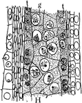
The cross section of a mature anther lobe in the stages of "formation of anthers and pollen grains or microspores of Silphium." -Stevens, 1916

Microspore Anther Lobe

The cross section of a mature anther lobe in the stages of "formation of anthers and pollen grains or…
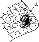
The archesporium in the stages of "formation of anthers and pollen grains or microspores of Silphium." -Stevens, 1916

Microspore Archesporium

The archesporium in the stages of "formation of anthers and pollen grains or microspores of Silphium."…
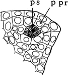
The primary sporogenous cell (ps) and the primary parietal layer (ppr) in the stages of "formation of anthers and pollen grains or microspores of Silphium." -Stevens, 1916

Microspore Cells

The primary sporogenous cell (ps) and the primary parietal layer (ppr) in the stages of "formation of…
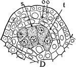
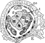
The sporogenous cells (s), tapetum (t), two parietal layers (oo) in the stages of "formation of anthers and pollen grains or microspores of Silphium." -Stevens, 1916

Microspore Cells

The sporogenous cells (s), tapetum (t), two parietal layers (oo) in the stages of "formation of anthers…

The sporogenous cells (s), tapetum (t), two parietal layers (oo) in the stages of "formation of anthers and pollen grains or microspores of Silphium." -Stevens, 1916

Microspore Cells

The sporogenous cells (s), tapetum (t), two parietal layers (oo) in the stages of "formation of anthers…
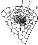
The inner layer (i), outer layer (o), sporogenous cells (s) in the stages of "formation of anthers and pollen grains or microspores of Silphium." -Stevens, 1916

Microspore Layers

The inner layer (i), outer layer (o), sporogenous cells (s) in the stages of "formation of anthers and…

The pollen grains beginning to germinate in the stages of "formation of anthers and pollen grains or microspores of Silphium." -Stevens, 1916

Microspore Pollen Grains

The pollen grains beginning to germinate in the stages of "formation of anthers and pollen grains or…

"Diagrammatic representation of a single palisade cell, with chloroplasts lining the walls." -Stevens, 1916

Palisade Cell

"Diagrammatic representation of a single palisade cell, with chloroplasts lining the walls." -Stevens,…

"Diagram to show the activities going on in a palisade cell. The arrows from the chloroplasts into the cell cavity indicate movement of photosynthesized product. The thickness of the arrow shafts under "Light" indicate the relative light intensities." -Stevens, 1916

Palisade Cell

"Diagram to show the activities going on in a palisade cell. The arrows from the chloroplasts into the…
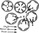
"Diagram to show the intake of carbon dioxide by the palisade cells from the intercellular spaces, the absorption by the chloroplasts of water from the cell-sap, and the passage of food from the chloroplasts into the cell-sap." -Stevens, 1916

Palisade Cell Intake

"Diagram to show the intake of carbon dioxide by the palisade cells from the intercellular spaces, the…
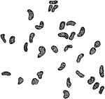
"Starch grains from the palisade cells of a Euphorbia leaf." -Stevens, 1916

Euphorbia Palisade Cells

"Starch grains from the palisade cells of a Euphorbia leaf." -Stevens, 1916

"Diagram to show the effect of different portions of the spectrum on photosynthesis. a to F, different regions of the spectrum from red to blue. A filamentous alga lies across these, and bacteria are collecting about the alga, with greatest frequency in the red between B and C, indicating the greatest evolution of oxygen there." -Stevens, 1916

Photosynthesis

"Diagram to show the effect of different portions of the spectrum on photosynthesis. a to F, different…
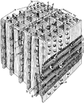
"Diagrammatic representation of a block of pine wood highly magnified. a, Early growth; b, late growth; c, intercellular space; d, bordered pit in tangential wall of late growth; m, f and e, bordered pit in radial wall of early growth from different points of view; h, row of medullary cells for carrying food; g, row of medullary ray cells for carrying water; k, thin place in radial wall of ray cells that carry food." -Stevens, 1916

Magnified Pine

"Diagrammatic representation of a block of pine wood highly magnified. a, Early growth; b, late growth;…

"Different stages in the development of a bordered pit. b, The original, thin, primary wall; a, the overhanging border formed as the wall thickened. B, Thickening of the wall has continued and extended the border; the primary wall has thickened at c, forming the torus. C, the border and the torus are finished." -Stevens, 1916

Pit Development

"Different stages in the development of a bordered pit. b, The original, thin, primary wall; a, the…

"Showing method of association of paternal and maternal chromosomes, at A in all vegetative nuclei, and B, C, D, their manner of separation in all vegetative nuclear divisions. Showing also manner of association of paternal and maternal chromosomes at E in early prophase of spore grandmother cells and the manner of their separation during the division of the grandmother-cell nucleus at F and G. Showing also the longitudinal division of the chromosomes in the spore mother cells in H, and their separation to form the spore nuclei in I, two of the four spores being purely paternal and two purely maternal." -Stevens, 1916

Plant Reproduction

"Showing method of association of paternal and maternal chromosomes, at A in all vegetative nuclei,…
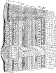
"Diagram showing additions to the primary tissues through the activity of the cambium and phellogen or cork cambium." -Stevens, 1916

Plant Tissues

"Diagram showing additions to the primary tissues through the activity of the cambium and phellogen…

"Diagram showing the relation of this year's leaves to the wood of the current year." -Stevens, 1916

Plant Tissues

"Diagram showing the relation of this year's leaves to the wood of the current year." -Stevens, 1916

"Apex of root hair flattened out over and imbedding soil particles." -Stevens, 1916

Plant Root Hair

"Apex of root hair flattened out over and imbedding soil particles." -Stevens, 1916

"A single root hair on a large scale, showing that it is an outgrowth of an epidermal cell, and the fact that it possesses a living protoplast and large vacuole filled with cell-sap and traversed by cytoplasmic strands. The nucleus is near the apex of the hair." -Stevens, 1916

Plant Root Hair

"A single root hair on a large scale, showing that it is an outgrowth of an epidermal cell, and the…

"Cross section of a root in the region of the root hairs." -Stevens, 1916

Plant Root Hairs

"Cross section of a root in the region of the root hairs." -Stevens, 1916

"Laticiferous vessels from the cortex of root of Scorozonora hispanica...B, smaller portion." -Stevens, 1916

Root Vessels

"Laticiferous vessels from the cortex of root of Scorozonora hispanica...B, smaller portion." -Stevens,…
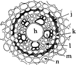
"Portion of a cross section of a root of Allium ascalonicum. h, large central, tracheal tube; i, xylem, and l, phloem portion of the vascular bundle; n, cortex cells; 1, endodermis with thin-walled cell at k to admit passage of materials; m, pericycle or pericambium." -Stevens, 1916

A. Ascalonicum Root

"Portion of a cross section of a root of Allium ascalonicum. h, large central, tracheal tube; i, xylem,…

"Diagram showing the progressive development of the skeletal tissues from the apex toward the base of the stem." -Stevens, 1916

Plant Skeletal Tissues

"Diagram showing the progressive development of the skeletal tissues from the apex toward the base of…

"Sphaero-crystals of unilin from tuber of Dahlia variabilis. A, precipitated from an aqueous solution; B, precipitated within the cells by long standing in alcohol." -Stevens, 1916

Sphaero-Crystals

"Sphaero-crystals of unilin from tuber of Dahlia variabilis. A, precipitated from an aqueous solution;…