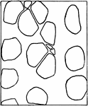
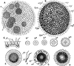
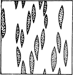
Starch
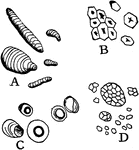
"Starch from different sources. A, curcuma starch; B, corn starch; C, tapioca starch; D, rice starch,…
Starch Grain Striations
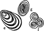
"Showing concentric and eccentric striations of starch grains. e, potato starch eccentrically striated;…
A. Capreolata Unusual Stem Growth
"Diagram showing some types of unusual growth in thickness. A, cross section through a four-year-old…
Bauhinia Unusual Stem Growth
"Diagram showing some types of unusual growth in thickness...B, cross section of stem of species of…
G. Scandens Unusual Stem Growth
"Diagram showing some types of unusual growth in thickness...C, portion of a cross section of stem of…
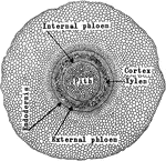
P. Galapageium Stem Tissue
The cross section of the stem of Pisdium Galapageium, which carries very little water.
Water Cress Stem Tissue
The cross section of the stem of water cress, which carries very little water.
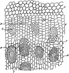
Yellow Poplar Stem Tissue
The cross section of the stem of yellow poplar, which carries large amounts of water.
A. Pedaium Stem
"Adianium pedatum: transverse section of stem, showing the amphiphloic siphonostele." -Stevens, 1916
C. Viticella Stem
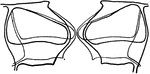
"Diagram showing the course of the vascular bundles in a stem of Clematis viticella. Median bundles…
Cerastium Stem
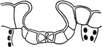
"B, diagram of vascular bundles in external view and in cross section of the stem of Cerastium. The…
D. Marginata Stem
"Portion of a cross section throughout the stem of Dracaena marginata. P, parenchyma of cortex. V, meristematic…
G. Pubescens Stem
"Gleichenia pubescens: transverse section of stem, showing the protostele." -Stevens, 1916
Ivy Stem
"Schizogenous resin duct in the young stem of ivy (Hedera helix), as seen in cross section. A, early,…
Juncus Stem
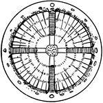
"A, cross section of stem of Juncus...the black lines traversing the section are really chains of cells…
P. Aquilina Stem
"Pteris aquilina: transverse section of stem, showing the polystele." -Stevens, 1916
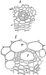
Palm Stem
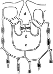
"Cross section of a portion of palm stem. e, xylem; f, phloem portions of vascular bundle; g, sclerenchyma…
Palm Stem
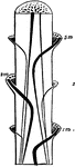
"A, diagram of course of vascular bundles in a palm stem; 1m, 2m, 3m, bundles from the median portions…
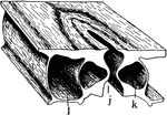
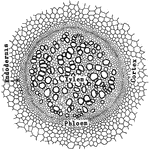
Stoma
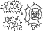
"A typical stoma in cross section and surface view combined. k, guard cell; j, the gap or stoma between…
Stoma Guard Cells
"A, diagram showing relative position of the guard cells in cross section in the open and closed positions;…
Depressed Stoma
"A, depressed stoma of the under side of a leaf of Amherstia nobilis." -Stevens, 1916
Depressed Stoma
"B, depressed stoma of Hakea suaveolens. g stands beneath the guard cells; d, outer, and e inner, cavities."…
Stomata Formation
"B and C, early stages in the formation of stomata; at s, mother cells of guard cells are shown. D,…
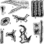
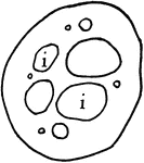
Stone Cells
"Stone cells from different sources. 1, from coffee; 2, 3, and 4, from stem of clove; 5 and 6, from…
Stone Cells 1
First developmental stage of stone cells: "1, in the primary meristem condition." -Stevens, 1916
Stone Cells 2
Second developmental stage of stone cells: "2, the cells have enlarged and the walls have begun to thicken…
Stone Cells 3
Third and final developmental stage of stone cells: "3, The walls are completed. The primary wall is…
I. Balsamina Storage Tissues
"Storage tissues of the cotyledon of Impatiens Balsamina. A, from the resting seed, and B, from a germinating…
Stored Food
"Diagram to show path of stored food upward through the tracheal tubes, and through the phloem portion…
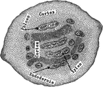
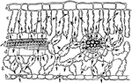
Marchantia Thallus
"Cross section through the thallus of Marchantia. j, stoma leading into a relatively large air-chamber…
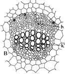
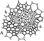
Collateral Vascular Bundle
Types of vascular bundles: "B, the collateral type, with phloem, h, standing in front of the xylem,…
Concentric Vascular Bundle
Types of vascular bundles: "A, the concentric type, with xylem, k, surrounding the phloem, h." -Stevens,…
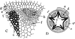
Radial Vascular Bundle
Types of vascular bundles: "C, a portion of the radial type, shown complete in D, where the part outlined…
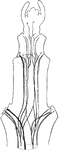
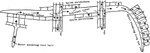
Vascular Bundles
"Diagram showing how the vascular bundles anastomose around the medullary rays. The gaps represent the…
Plant Vein
"Diagram indicating the succession of the conducting tissues of a vein from the base toward the apex.…
Volvox Globator
"Volvox globator. A, entire colony, enclosing several daughter-colonies; B, the same during sexual maturity;…
Leaf Water Flow
"Semi-diagrammatic cross section of a leaf showing by arrows how the water passes from the tracheal…
Leaf Water Flow
"Diagram to show the path of the water as it rises to, and escapes from, the leaves." -Stevens, 1916
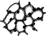
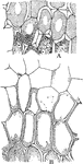
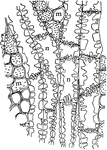
Grapevine Wood
"Tangential section through the wood of grapevine. m, cells of medullary ray, and n, of xylem parenchyma,…
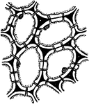
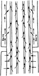
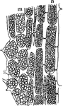
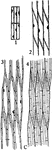
L. Tulipifera Wood
"Tangential section of wood of Liriodendron tulipifera, showing frequency of contact of medullary rays…
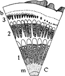
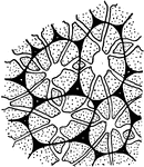
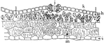
Oak Wood
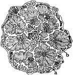
"Outline of tangential section of wood of oak, to show frequency of medullary rays. The section is 1…
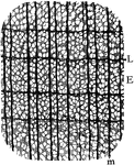
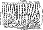
Yellow Poplar Wood
"Cross section of yellow poplar wood. E, early; L, late growth; m, medullary ray." -Stevens, 1916
Yellow Poplar Wood
"Showing pitted connections between medullary rays and xylem parenchyma, and between contiguous xylem…
Xylem Development 1
"Stages in the development of the elements of the xylem. A, progressive steps in the development of…
Xylem Development 2
Stages in the development of the elements of the xylem. "B, stages in the formation of tracheids from…
Xylem Development 3
Stages in the development of the elements of the xylem. "C, steps in the development of wood fibers…
Xylem Development 4
Stages in the development of the elements of the xylem. "D, steps in the formation of wood parenchyma…