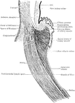
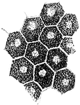
Lines for the detection of astigmatism. "The refracting surfaces of the eye acting together are equivalent in refracting power to a single, spherical surface of fairly short curvature. Frequently, however, the result is not the same as would be given by a perfect spherical surface, owing to inequalities in the curvature of the eye. In one direction the curvature may be greater than that at right angles to it. This tendency to a cylindrical form is called astigmatism. It interferes with the formation of perfect images and sometimes leads to serious eye strain in the effort to better the vision. Astigmatism may be detected by looking at black lines radiating from a point or at fine black concentric circles. Portions of the liens or circles appear gray and others black; the gray portions are out of focus. This defect is corrected by proper cylindrical glasses which equalize the curvature of the eye" — Newell, 1900

Detection of Astigmatism

Lines for the detection of astigmatism. "The refracting surfaces of the eye acting together are equivalent…

A. A cone and two rods from the human retina (modified from Max Schultze); B. Outer part of rod separated into discs.

Cones and Rods of Retina

A. A cone and two rods from the human retina (modified from Max Schultze); B. Outer part of rod separated…
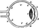
A section of the human eye.

Eye

A section of the human eye.

"a, sclerotic membrane; b, cornea; d, retina; o, optic nerve; v, vitreous humor." -Comstock 1850

Eye

"a, sclerotic membrane; b, cornea; d, retina; o, optic nerve; v, vitreous humor." -Comstock 1850
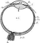
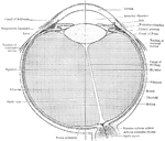
"Diagram of the eye. C., Cornea; a.h., aqueous humour; c.b., ciliary body; l., lens; I., iris; Sc., sclerotic; Ch., choroid; R., retina; v.h., vitreous humour; y.sp., yellow spot; n., optic nerve." -Thomson, 1916

Eye

"Diagram of the eye. C., Cornea; a.h., aqueous humour; c.b., ciliary body; l., lens; I., iris; Sc.,…
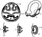
"Development of the eye. 1. Section through first embryonic vesicle, showing outgrowth of optic vesicles (op.v.) to meet the skin; j.b., thalamencephalon; G., the gut. 2-4. Sections illustrating the formation of the lens (l.) from the skin, and the modification of the optic vesicle into an optic cup; R., retina; v.h., vitreous humour. 5. External aspect of embryonic eye; l., lens." -Thomson, 1916

Eye Development

"Development of the eye. 1. Section through first embryonic vesicle, showing outgrowth of optic vesicles…
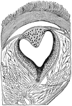
Longitudinal section through the pineal eye of a lizard. The eye is located in the middle of the dorsal side of the head and is covered by the translucent scaled. The outer wall of the eye vesicle is thickened to form a lens, while the inner pigmented wall is the retina from which the nerve proceeds.

Eye of a Lizard

Longitudinal section through the pineal eye of a lizard. The eye is located in the middle of the dorsal…
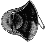
A section of the eye seen from within. Labels: 1, The divided edge of the three coats. 2, The pupil. 3, The iris. 4, The ciliary processes. 5, The scolloped border of the retina.

A Section of the Eye Seen from Within

A section of the eye seen from within. Labels: 1, The divided edge of the three coats. 2, The pupil.…

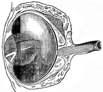
The eye of a common owl. The length from the anterior to posterior diameter lengthened.

Bird Eye

The eye of a common owl. The length from the anterior to posterior diameter lengthened.

"If the cornea is too convex, or prominent, the image will be formed before it reaches the retina, for the same reason, that of two lenses, that which is most convex will have the least focal distance." -Comstock 1850

Cornea too Convex on Eye

"If the cornea is too convex, or prominent, the image will be formed before it reaches the retina, for…
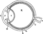
Plan of the eye seen in section. Labels: A, The Sclerotic Coat; B, The Choroid Coat; C, The Retina; D, The Optic Nerve; E, The Cornea; F, The Aqueous Humor; G, The Iris; H, The Ciliary Processes; I. The Crystalline Lens; K, The Vitreous Humor.

Diagram of the Eye

Plan of the eye seen in section. Labels: A, The Sclerotic Coat; B, The Choroid Coat; C, The Retina;…

"Diagram illustrating the Manner in which the Image of an Object is inverted on the Retina." — Blaisedell, 1904

Diagram of the Eye

"Diagram illustrating the Manner in which the Image of an Object is inverted on the Retina." — Blaisedell,…

The eye of a fish. The anterior to posterior diameter has been decreased.

Fish Eye

The eye of a fish. The anterior to posterior diameter has been decreased.

"Showing how the image of an object which is seen is formed on the retina of the eye." —Croft 1917
Eye Focusing on Object

"Showing how the image of an object which is seen is formed on the retina of the eye." —Croft 1917

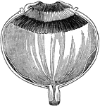
Section of the eye magnified, showing the ciliary processes, the pigmentum nigrum, the retina, and the choroid coat.

Section of the Eye

Section of the eye magnified, showing the ciliary processes, the pigmentum nigrum, the retina, and the…
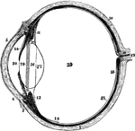
The left eyeball in horizontal section from before back. Labels: 1, sclerotic; 2, junction of sclerotic and cornea; 3, cornea; 4, 5, conjunctival membrane; 7, ciliary muscle; 10, choroid; 11, 13, ciliary processes; 14, iris; 15, retina; 16, optic nerve; 17, artery entering retina; 18, fovea centralis; 19, region where sensory part of retina ends; 26, 27, 28, are placed on the lens; 28 , suspensory ligament placed around lens; 29, vitreous humor; 30, aqueous humor in anterior chamber.

Left Eyeball in Horizontal Section

The left eyeball in horizontal section from before back. Labels: 1, sclerotic; 2, junction of sclerotic…
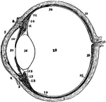
Diagram of a horizontal section through left eyeball and optic nerve.

Eyeball and Optic Nerve

Diagram of a horizontal section through left eyeball and optic nerve.

The left eyeball in horizontal section from before back. Labels: 1, sclerotic; 2, junction of sclerotic and cornea; 3, cornea; 4, 5, conjunctiva; 7, ciliary muscle; 10, choroid; 11, 13, ciliary processes; 14, iris; 15, retina; 16, optic nerve; 17, artery entering retina in optic nerve; 18, fovea centralis; 19, 20, region where sensory part of retina ends; 22, suspensory ligament; 24, the anterior part of the hyaloid membrane; 26, the lends; 29, vitreous humor; 30, aqueous humor.

The Eyeball in Horizontal Section

The left eyeball in horizontal section from before back. Labels: 1, sclerotic; 2, junction of sclerotic…
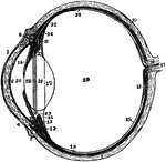
The left eyeball in horizontal section from before back. Labels: 1, sclerotic; 2, junction of sclerotic and cornea; 3, cornea; 4,5, conjunctiva; 6, posterior elastic layer of cornea; 7, ciliary muscle; 10, choroid; 11,13, ciliary processes; 14, iris; 15, retina; 16, optic nerve; 17, artery entering retina in optic nerve; 18, fovea centralis; 19, region where sensory part of retina ends; 22, suspensory ligament; 23 is placed in the canal of Petit, and the line from 25 points to it; 24, the anterior part of the hyaloid membrane; 26, 27, 28, are placed on the lends; 28 points to the line of attachment around it of the suspensory ligament; 29, vitreous humor; 30, anterior chamber of aqueous humor; 31, posterior chamber of aqueous humor.

Section of Left Eyeball

The left eyeball in horizontal section from before back. Labels: 1, sclerotic; 2, junction of sclerotic…

Section through ciliary region of eyeball.

Section Through Eyeball

Section through ciliary region of eyeball.

"A representation of the manner in which the image is formed in the eye, when the cornea or crystalline lens is flattened. The perfect image would be produced at m m, behind the retina, and, of course, beyond the point necessary to secure perfect vision." —Wells, 1857

Flattened Eye

"A representation of the manner in which the image is formed in the eye, when the cornea or crystalline…
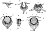
"Diagrams showing some of the stages in the increasing complexity of the simple eye in Invertebrates. A, simple pigment spot in epithelium having nerve-endings associated with pigment cells (as in some medusae); B, pigment cells in a pit-like depression (as in Patella); C, with pin-hole opening and vitreous humor in cavity (as in Trochus); D, completely closed pit, with lens and cornea (as in Triton and many other Mollusks); E, pigment area elevated instead of depressed, lens of thickened cuticula (as in the Medusa, Lizzia); F, retinal cells more highly magnified. ep., epidermis; f, nerve fibre; l, lens; op, optic nerve; p, pigment cells; r, retina; v.h., vitreous humor." -Galloway, 1915

Invertebrate Simple Eye

"Diagrams showing some of the stages in the increasing complexity of the simple eye in Invertebrates.…

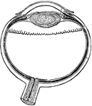
A convex lens, bends the ray of light which pass through it, so that they meet at a point called a focus. The crystalline lens converges the rays of light which enter the eye, and brings them to a focus on the retina. The healthy lens has a power of changing its convexity so as to adapt itself to near and to distant objects.

The Use of the Crystalline Lens

A convex lens, bends the ray of light which pass through it, so that they meet at a point called a focus.…

The muscles of the eyeball, the view being taken from the outer side of the right orbit.

Eye Muscles

The muscles of the eyeball, the view being taken from the outer side of the right orbit.
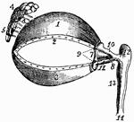
1, cartilage of the upper eyelid; 2, its lower border, showing the openings of the Meibomian glands; 3, the cartilage of the lower eyelids, also showing its border the openings of the Meibomian glands; 4, 5, the lachrymal glands; 6, its ducts; 7, the plica semilunaris; 8, the caruneula lachrymal canals; 10,11, the superior and inferior lachrymal canals; 12, the lachrymal sac; 13, the nasal duct, terminating at 14 in the lower meatus of the nose.

Eye Muscles

1, cartilage of the upper eyelid; 2, its lower border, showing the openings of the Meibomian glands;…

"A representation of the manner in which the image is formed upon the retina in the perfect eye. The curvature of the cornea, s s, and of the crystalline lens, c c, is just sufficient to cause the rays of light proceeding from the image, e e, to converge the right focus, m, upon the retina." —Wells, 1857

Perfect Eye

"A representation of the manner in which the image is formed upon the retina in the perfect eye. The…

Pigment epithelium of human retina (viewed from the surface).

Pigment Epithelium of Retina

Pigment epithelium of human retina (viewed from the surface).

"Diagrammatic section through the eyeball. xx, optic axis; k, nodal point." —Martin, 1917

Retina

"Diagrammatic section through the eyeball. xx, optic axis; k, nodal point." —Martin, 1917

"Diagram illustrating the points at which incident rays meet the retina. xx, optic axis; k, first nodal point; k', second nodal point; b, point where the image of B would be formed, were the eye properly accommodated for it; a, the retinal point where the image of A would be formed." —Martin, 1917

Retina

"Diagram illustrating the points at which incident rays meet the retina. xx, optic axis; k, first nodal…

A diagram showing how an image is formed upon the retina by the crystalline lens.

A Diagram of the Retina and Crystalline Lens

A diagram showing how an image is formed upon the retina by the crystalline lens.
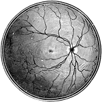
The right retina as it would be seen if the front part of the eyeball with the lens and vitreous humor were removed. The white disk to the right marks the entry of the optic nerve (blind spot); the lines radiating from this are the retinal arteries and veins. The small central dark patch is the yellow spot, the region of most acute vision.

Retina Blind Spot

The right retina as it would be seen if the front part of the eyeball with the lens and vitreous humor…

Diagram showing the sustentacular fibers of the retina fiber basket above the external lumbar membrane; m, nucleus of the fiber; r, base of the fiber.

Sustentacular Fibers of the Retina Fiber Basket

Diagram showing the sustentacular fibers of the retina fiber basket above the external lumbar membrane;…

A flattened shape of the globe of the eye causes farsightedness and an elongated shape of the globe of the eye causes nearsightedness. The diagram illustrates the position of the retina in these conditions. Labels: B, natural sight; G, farsightedness; C, nearsightedness.

The Position of the Retina in Near and Far Sight

A flattened shape of the globe of the eye causes farsightedness and an elongated shape of the globe…

A section through the retina from it anterior inner surface (1) in contact with the hyaloid membrane, to its outer (10) in contact with the choroid. 1, internal limiting membrane; 2, nerve-fiber layer; 3, nerve cell layer; 4, inner molecular layer; 5, inner granular layer; 6, outer molecular layer; 7, outer granular layer; 8, external limiting membrane; 9, rod and cone layer; 10, pigment cell layer.

Section of Retina

A section through the retina from it anterior inner surface (1) in contact with the hyaloid membrane,…
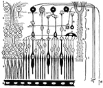
The retina of a human eye stretched out vertical.

Eye Retina

The retina of a human eye stretched out vertical.

Diagram of the retina, aka percipient layer of the eye. 1: inner limiting membrane, which is next to the vitreous humor in the eye cavity; 2: a branch of a divided optic nerve; 3: ganglion cell layer; 4: inner nerve-fiber layer; 5: layer of bipolar ("inner granular") cells; 6: outer layer of nerve-cells; 7: layer of visual (outer granular) cells; 8: outer limiting membrane; 9: long and thing rods and short and broad cones; 10: pigment layer; 11: tangential cells; 12: bipolar cells; 13: amacrine cell; 14: centripetal optic nerve fibers; 15: centrifugal optic nerve fibers; 16: Muller's supporting cells. I, II, III: three areas of nerve cells in the retina.

Eye Retina

Diagram of the retina, aka percipient layer of the eye. 1: inner limiting membrane, which is next to…

Formation of an image in the retina.

Image Formation in the Retina

Formation of an image in the retina.

Section through the macula lutea and fovea centralis of the human retina. Labels: a, fovea; b, descent of the macula towards fovea. The numbers indicate the layers of the retina: 1, membrana limitans interna; 2, optic nerve fibers; 3, layer of ganglionic corpuscles; 4, inner molecular layer; 5, inner nuclear layer; 6, intergranular, internuclear, or outer molecular layer. 7, external nuclear layer; 8, membrana limitans externa; 9, layer of rods and cones; 10, pigment-cell layer.

Layers of the Retina

Section through the macula lutea and fovea centralis of the human retina. Labels: a, fovea; b, descent…

Diagram showing the nervous elements of retina. Labels: 1, nerve fiber to ganglion cell; 2, processes of ganglion cell going outwards; 3, nerve fiber passing direct to granule in inner nuclear layer; 4, process of ganglion cell to granule; 5, fiber from cone-granule breaking up into fibrils which are connected with rods and cone fibrils.

Nervous Elements of the Retina

Diagram showing the nervous elements of retina. Labels: 1, nerve fiber to ganglion cell; 2, processes…

Diagram showing relations of the neurons and sensory epithelium in the retina. labels: E, epithelial layer of nucleated rode and cone cells, rods being directed towards choroid coat of retina; N1, neurons of first series receiving by their dendritic impulses from the rod and cone cells and transmitting them by their axons to N2, the neurons of the second series. The axons of the neurons of the second series pass along the inner surface of the retina to the blind spot, where they unite to form the optic nerve.

Neurons and Sensory Epithelium in the Retina

Diagram showing relations of the neurons and sensory epithelium in the retina. labels: E, epithelial…
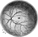
The posterior half of the retina of the left eye, viewed from before; s, the cut edge of the sclerotic coat; ch, the choroid; r, the retina; in the interior at the middle the macula lutea with the depression of the fovea centralis is represented by a slight oval shade; towards the left side the light spot indicates the colliculus or eminence at the entrance of the optic nerve, from the center of which the arteria centralis is seen spreading its branches into the retina, leaving the part occupied by the macula comparatively free.

Posterior Half of the Retina

The posterior half of the retina of the left eye, viewed from before; s, the cut edge of the sclerotic…

Diagrammatic section of the human retina.

Section of Retina

Diagrammatic section of the human retina.

Perpendicular section of mammalian retina. Labels: A, layer of rods and cones; B, outer nuclear layer; C, outer molecular layer; D, inner nuclear layer; E, inner molecular layer; F, ganglionic layer; G, stratum opticum; r, rods; c, cones, r.g, rod granules; c.g, cone granules; r.b, rod bipolars; c.b, cone bipolars; c.r, vontact of rod bipolars with the internal arborisation of cone bipolars; ar', internal arborisation of rod bipolars; c.n, centrifugal nerve fiber; h, horizontal cells; s.s, stratified spongioblasts; d.s, diffuse spongioblasts; s.g, stratified ganglion cell; M, sustentacular fiber of Muller.

Section of Retina

Perpendicular section of mammalian retina. Labels: A, layer of rods and cones; B, outer nuclear layer;…

Structure of the human retina, showing the various layers.

Structure of the Retina

Structure of the human retina, showing the various layers.

Next to the choroid and comprising about 1/4 the entire thickness of the retina is a multitude of transparent, colorless, microscopic rods (a) evenly arranged and packed side by side, like the seeds in the disk of a sunflower. Among them at regular intervals, are interspersed the cones (b). Delicate nerve fibers pass from the ends of the rods and cones, each expanding into a granular body (c) thence weaving a mesh (d) and again expanding into the granules (f). Last is a layer of fine nerve fibers (g) and gray, ganglionic cells (h) like the gray matter of the brain, whence filaments extend into the fibers of the optic nerve (i).

The Structure of the Retina

Next to the choroid and comprising about 1/4 the entire thickness of the retina is a multitude of transparent,…

A section of the retina, choroid, and part of the sclerotic. Labels: a, membrana limitans interna; b, nerve-fiber later traversed by Muller's sustentacular fibers (of the connective-tissue system): c, ganglion-cell layer; d, molecular layer; e, internal nuclear layer; f, internuclear layer; g, external nuclear layer; h, membrana limitans externa, running along the lower part of i, the layer of rods and cones; k, pigment cell layer; lm, internal and external vascular portions of the choroid, the first containing capillaries, the second larger blood-vessels, cut in transverse section; n, sclerotic.

Structure of the Retina

A section of the retina, choroid, and part of the sclerotic. Labels: a, membrana limitans interna; b,…
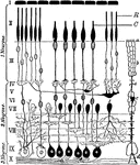
Diagram of the structure of the human retina. Labels: I, pigment layer; II, rod and cone layer; R, rods; C, cones; III-IX, intraretinal nerve-elements; X, axons which pass to optic nerve.

Retinal Structure

Diagram of the structure of the human retina. Labels: I, pigment layer; II, rod and cone layer; R, rods;…

"Diagrams illustrating two stages in the development of the vertebrate eye. A, showing the relation of the ectoderm, the brain vesicle, and the optic vesicle. The right side of the figure shows a later stage than the left. B, later stage, showing the lens, eye-ball and retina in position. b.v., brain vesicle formed by the invagination oof the ectoderm (ect.); l, lens; mes., mesodermal tissue; o.n., optic nerve; o.s., optic stalk; o.v., optic vesicle, a portion of the brain vesicle; r, retinal layer; v.h., interior of eye-ball which comes to contain the vitreous humor." — Galloway

Vertebrate Eye

"Diagrams illustrating two stages in the development of the vertebrate eye. A, showing the relation…