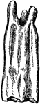
Scalpriform, Left Lower Incisor of a Beaver
Close-up illustration of scalpriform incisor of a beaver. It is "chisel-shaped; having the character…
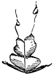
Structure of Canine Tooth
Vertical section of canine tooth to illustrate the various parts and structures.
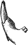
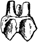
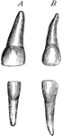
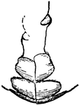
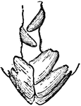
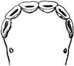
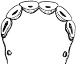
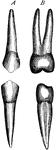
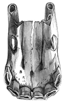
Temporary Canine
Canine teeth of left side, labiial (A) and lateral (B) aspects. C, temporary canines.
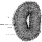
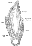
Dental Sac and Pulp
Vertical transverse section of the dental sac and pulp of a kitten. Labels: a, dental papilla or pulp;…
Dentine and Cement
Section of a portion of the dentine and cement from the middle of the root of an incisor tooth. Labels:…
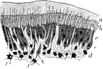
Deposition of Dentine
Part of section of developing tooth of a young rat, showing the mode of deposition of the dentine. Labels:…
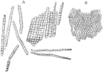
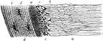
Enamel Fibers
Enamel fibers. A, fragments and single fibers of the transversely striated enamel, isolated by the action…
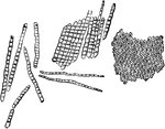
Enamel Prisms
Enamel prisms. A, Fragments and single fibers of the enamel isolated by the action of hydrochloric acid;…
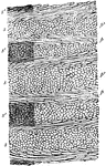
Longitudinal Section of Enamel
Longitudinal section of enamel, treated with acid, showing disposition of ranges of enamel prisms (p,…
Horse Molar
"Side view of second upper molar tooth of Anchitherium (brachyodont form)." — Encyclopedia Britannica,…
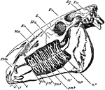
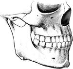
Horse Skull
"Side view of skull of horse, with the bone removed so as to expose the whole of the teeth. PMx, premaxilla;…
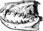
Hyaenodon Leptorhynchus
"Dentition of Hyaenodon leptorhynchus. The posterior molar is concealed behind the penultimate tooth."…
Jaw Showing Roots of Teeth
Horizontal section through both the upper and lower jaws to show the roots of the teeth. The sections…
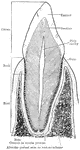
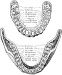
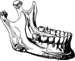
Lower Jaw with Teeth
Right half of lower jaw, with the corresponding teeth. The letters and numbers point to the various…
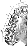
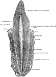
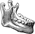
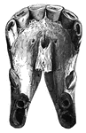
Upper Jaw with Teeth
Right half of upper jaw (from below), with the corresponding teeth. The letters and numbers point to…
Section of Tooth of Typical Labyrinthodont
This illustration shows a section of a tooth of a typical Labyrinthodont. Labyrinthodont, or Stegocephali…

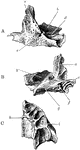
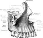
Ossification of Superior Maxilla
Ossification of superior maxilla. A, outer side. B, inner side. C, under side. Labels: a, nasal process;…
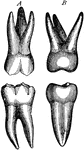
Comparison of the Molar Teeth of a Human, Horse, and Dog
The molar teeth of a human, horse and dog. The first image to the left in a molar tooth of a horse.…
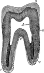
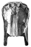
Structure of a Molar
Longitudinal section (A) and transverse section (B) of a human molar tooth. Labels: c, cement; d, dentine;…
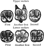
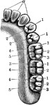
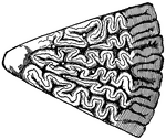
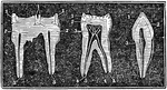
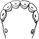
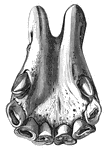
Surface of Molar
Triturating surfaces of molar teeth of right side. The upper margin of the figures corresponds to the…
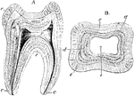
Vertical Section of a Molar
Vertical section of a molar tooth. Labels: a, enamel of the crown, the line of which indicate the arrangement…

Savart wheel
"The Savart wheel consists of a heavy metal toothed wheel that may be put in rapid revolution by pulling…
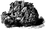
Dogtooth Spar
"...a variety of calcite, crystallizing in scalenohedral forms; so named from a fancied resemblance…
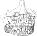
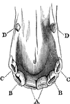
Teeth
To show the relation of the upper to the lower teeth when the mouth is closed. The manner in which a…
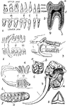
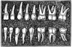
Teeth of Man and Several Animal Species
1. Dentition (teeth) of man. 2. Dentition of hyena. 3. Dentition of pig. 4. Dentition of Patagonian…
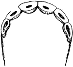
The Adult Teeth
The adult teeth. Labels: 1, 2, The cutting teeth (incisors). 3, Eyetooth (cuspid). 4,5, Small grinders…
Development of Permanent Teeth
Well formed jaws, from which the alveolar plate has been removed to expose the developing permanent…
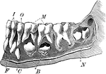
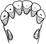
Emergence of Adult Teeth
The teeth of a 6 and a half year old child. Label: I, the incisors; O, the canine; M, the molars; the…
!["A Tooth is one of the hard bodies of the mouth, attached to the skeleton, but not forming part of it and developed from the dermis or true skin. True teeth consist of one, two, or more tissues differing in their chemical composition and in their microscopical appearances. Dentine, which forms the body of the tooth, and 'cement,' which forms its outer crust, are always present, the third tissue, the 'enamel,' when present, being situated between the dentine and cement. The incisors, or cutting teeth, are situated in front. In men there are two of these incisors in each side of each jaw. The permanent incisors, molars, and premolars are preceded by a set of deciduous or milk teeth, which are lost before maturity, and replaced by the permanent ones. The canines come next to the incisors. In man there is one canine tooth in each half-jaw. The premolars (known also as bicuspids and false molars) come next in order to the canines. In man there are two premolars in each half-jaw. The true molars (or multicuspids) are placed most posteriorly. In man there are three molars in each half-jaw, the posterior one being termed the wisdom tooth. The figures [in the illustration] refer to months after birth."—(Charles Leonard-Stuart, 1911)](https://etc.usf.edu/clipart/15200/15256/teeth1_15256_mth.gif)
First Teeth
"A Tooth is one of the hard bodies of the mouth, attached to the skeleton, but not forming part of it…
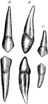
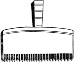
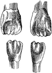
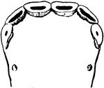
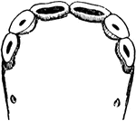
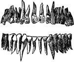
Incisor and Canine Horse Teeth
Incisor and canine teeth of a horse. A, front, B, lateral, and C, corner incisor; D, canine teeth.