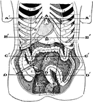
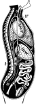
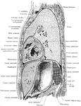
Abdomen Laid Open After Removal of Jejunum and Ileum
The abdomen viscera after the removal of the jejunum and ileum. The transverse colon is much more regular…
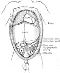
Abdomen of Fetus
The abdominal and thoracic viscera of a five months fetus. The large liver and large size if its left…
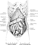
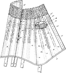
Abdomen Showing Displacement Caused by Corset
Abdomen of female showing displacement resulting from tight lacing. The liver is much enlarged, and…
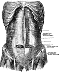
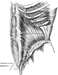
Muscles of the Abdomen
The muscles of the abdomen, showing the semilunar fold of Douglas. Viewed from in front.
Transverse Section of Abdomen
Diagrammatic transverse section of abdomen, to show the peritoneum on transverse tracing. A, at level…
Transverse Section of the Abdomen
A transverse section of the abdomen in the lumbar region showing abdominal muscles.
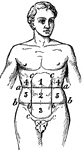
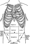
Abdominal
"In human anatomy, certain regions into which the abdomen is arbitrarily divided for the purpose of…
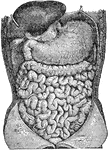
Abdominal Organs
Abdominal organs. Labels: 1, liver turned up; 2, gall bladder; 3, stomach; 4, large intestine; 5, small…
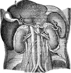
Abdominal Organs
Abdominal organs. Labels: 1, liver turned up; 2, gall bladder; 3, right kidney; 4, spleen; 5, left kidney.
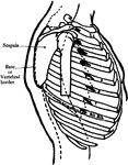
Abdominal Region
Showing the average position of the abdominal viscera with their surface markings. Labels: A, sterno-ensiform…
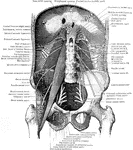
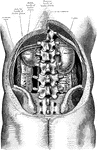
Muscles of the Abdominal Wall
View of the posterior abdominal wall to show the muscles and the nerves of the lumbo sacral plexus.
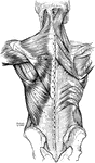
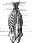
Back Muscles
Muscles of the back. On the left side is exposed the first layer; on the right side, the second layer…
A Side View of the Chest and Abdomen in Respiration
A side view of the chest and abdomen in respiration. Labels: 1, The cavity of the chest. 2, The cavity…
![Torso of the Statue known as Venus of Melos (left) and New York Fashion, 1898 (right). "Since abdomen and chest alternately expand and contract in healthy breathing, anything which impedes their free movement is to be avoided. The tight lacing which is still indulged [in 1900] by those who think a distorted form beautiful, seriously impedes one of the most important functions of the body, and leads not only to shortness of breath and an incapacity for muscular exertion, but in many cases to actual deformity or disease." — Newell, 1900.](https://etc.usf.edu/clipart/36200/36290/corset_36290_mth.gif)
Effect of Corset Use on Respiration
Torso of the Statue known as Venus of Melos (left) and New York Fashion, 1898 (right). "Since abdomen…
![Torso of the Statue known as Venus of Melos (left) and New York Fashion, 1898 (right). "Since abdomen and chest alternately expand and contract in healthy breathing, anything which impedes their free movement is to be avoided. The tight lacing which is still indulged [in 1900] by those who think a distorted form beautiful, seriously impedes one of the most important functions of the body, and leads not only to shortness of breath and an incapacity for muscular exertion, but in many cases to actual deformity or disease." — Newell, 1900.](https://etc.usf.edu/clipart/36200/36291/corset_36291_mth.gif)
Effect of Corset Use on Respiration
Torso of the Statue known as Venus of Melos (left) and New York Fashion, 1898 (right). "Since abdomen…
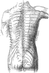
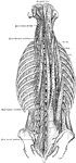
Posterior View of the Cutaneous Nerves of Trunk
The distribution of cutaneous nerves n the back of the trunk. On the left side the distribution of the…
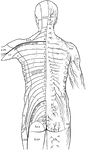
Distribution of Cutaneous Nerves on the Back
The distribution of the cutaneous nerves on the back of the trunk. On one side the distribution of the…
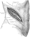
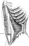
Surgical Incision to the Kidney
An incision in the right side above the kidney, showing a typical surgical approach to this organ, exposing…
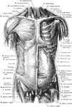
Anterior View of the Muscles of the Trunk
Superficial and deep muscles of the trunk. The sternocleidomastoid, pectoralis major, anterior portion…
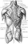
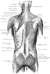
Posterior View of the Muscles of the Trunk
Superficial and deep muscles of the trunk. The latissimus dorsi and trapezius on the right side have…

View of Organs from the Side
The chief organs of the body from the side. Labels: a, arch of the aorta or main artery of the trunk;…
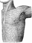
Lymphatics of the Shoulder
Lymphatics and lymphatic glands on the left side of the body and shoulder.
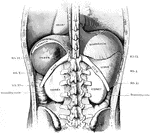
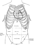
Diagram of Thoracic and Abdominal Regions
A diagram of the thoracic and abdominal regions. Labels: A, aortic valve; M, mitral valve; p, pulmonary…
Thoracic and Abdominal Regions
Diagram of the thoracic and abdominal regions. Labels: A, aortic valve; P, pulmonary valve; M, mitral…
Side View of the Thorax and Part of the Abdomen
Lateral, sagittal section through the left thorax and upper portion of abdomen, viewed from the left.…
Torso
The human torso. Labels: A, the heart; B, the lungs drawn aside to show the internal organs; C, the…
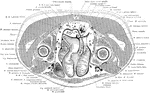
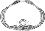
Cross Section of the Trunk above Pubic Symphysis
Section through the pelvis immediately above the pubic symphysis.
Cross Section of the Trunk above the Manubrium Sterni
Section immediately above the manubrium sterni.
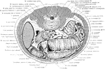
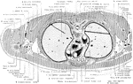
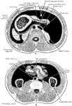
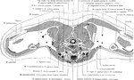
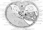
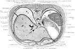
Cross Section of the Trunk Above Umbilicus
Section through the abdomen, an inch and a half above the umbilicus. The liver is unusually large in…
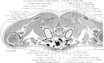
Cross Section of the Trunk at First Sacral Vertebra
Section through the pelvis at the level of the first sacral vertebra.
Cross Section of the Trunk at the Level of Junction of the Manubrium
Section at the level of the junction of the manubrium and corpus sterni, exposing the great vessels…
Cross Section Through the Trunk at the Neck
Section through the neck immediately above the shoulders.
Cross Section Through the Trunk at the Shoulders
Section through the thyroid gland and the upper borders of the shoulders.
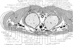
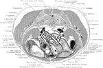
Cross Section of the Trunk Below Umbilicus
Section through the abdomen immediately below the umbilicus and the superior iliac crest.
Cross Section of the Trunk Exposing the Ventricles of the Heart
Section exposing the ventricles of the heart.
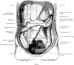
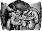
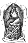
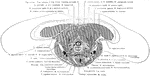
Trunk Showing Organs of Digestion
Diagram of the relations of the large intestine and kidneys, from behind.
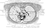
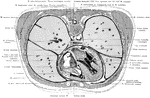
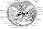
Cross Section of the Trunk through Costal Arch
Section passes through the lower portion of the costal arch, and through the hilus of the two kidneys.
Cross Section of the Trunk through the First Lumbar Vertebra
Section through first lumbar vertebra and one inch below ensiform process.
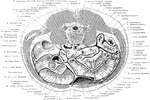
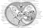
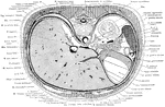
Cross Section of the Trunk through the Liver
Section through the body of the liver at the level of the arch of the seventh rib anteriorly.
Cross Section of the Trunk through Third Lumbar Vertebra
Section through the third lumbar vertebra and the inferior poles of the kidneys cutting the loop of…
Cross Section of the Trunk through Upper Kidney
Section through the upper pole of the left kidney at the level of the tip of the xiphoid process.
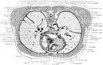
Cross Section of the Trunk Through the Inferior Portion of the Heart
Section through the inferior portion of the heart, exposing the dome of the diaphragm on the right side.
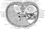
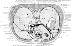
Cross Section of the Trunk Through the Liver and Stomach
Section through the liver and stomach, at the level of the xiphoid process.
Cross Section of the Trunk Through the Lungs
Section through the humeral heads and the apices of the lungs.