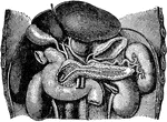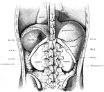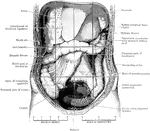
The abdomen viscera after the removal of the jejunum and ileum. The transverse colon is much more regular than usual. Both the liver and caecum extend lower down than normal. The subdivisions of the abdominal cavity are indicated by dark lines.

Abdomen Laid Open After Removal of Jejunum and Ileum

The abdomen viscera after the removal of the jejunum and ileum. The transverse colon is much more regular…
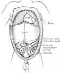
The abdominal and thoracic viscera of a five months fetus. The large liver and large size if its left lobe at this age should be noted.

Abdomen of Fetus

The abdominal and thoracic viscera of a five months fetus. The large liver and large size if its left…
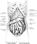
Abdomen of female showing displacement resulting from tight lacing. The liver is much enlarged, and extends on the left side to the ribs, where it was folded back on itself for over an inch. The pyloric end of the stomach and the beginning of the duodenum are quite superficial below the liver, and all the viscera are displaced downwards.

Abdomen Showing Displacement Caused by Corset

Abdomen of female showing displacement resulting from tight lacing. The liver is much enlarged, and…
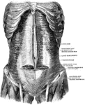
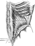
The muscles of the abdomen, showing the semilunar fold of Douglas. Viewed from in front.

Muscles of the Abdomen

The muscles of the abdomen, showing the semilunar fold of Douglas. Viewed from in front.
The transversalis, rectus, and pyramidalis muscles.

Muscles of the Abdomen

The transversalis, rectus, and pyramidalis muscles.

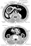
Diagrammatic transverse section of abdomen, to show the peritoneum on transverse tracing. A, at level of foramen of Winslow. B, lower down. In A note, one of the vasa brevia arteris passing to the stomach between the layers of the gastrosplenic omentum, and also the foramen of Winslow leading into the lesser sac which lies behind the stomach.

Transverse Section of Abdomen

Diagrammatic transverse section of abdomen, to show the peritoneum on transverse tracing. A, at level…
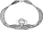
A transverse section of the abdomen in the lumbar region showing abdominal muscles.

Transverse Section of the Abdomen

A transverse section of the abdomen in the lumbar region showing abdominal muscles.
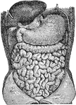
Abdominal organs. Labels: 1, liver turned up; 2, gall bladder; 3, stomach; 4, large intestine; 5, small intestine.

Abdominal Organs

Abdominal organs. Labels: 1, liver turned up; 2, gall bladder; 3, stomach; 4, large intestine; 5, small…
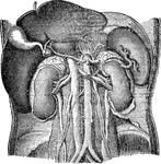
Abdominal organs. Labels: 1, liver turned up; 2, gall bladder; 3, right kidney; 4, spleen; 5, left kidney.
Abdominal Organs

Abdominal organs. Labels: 1, liver turned up; 2, gall bladder; 3, right kidney; 4, spleen; 5, left kidney.

Abdominal organs. Labels: 1, liver; 2, gall bladder; 3, spleen; 4, pancreas.

Abdominal Organs

Abdominal organs. Labels: 1, liver; 2, gall bladder; 3, spleen; 4, pancreas.

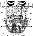
Showing the average position of the abdominal viscera with their surface markings. Labels: A, sterno-ensiform point; A', sterno-ensiform line; B, mid-epigastric point; B', mid-epigastric or transpyloric line; C, umbilical point; C', umbilical line; D, mid-hypogastric point; D', mid-hypogastric line; E, outer border of the right and left rectus abdominis; F, Monro's point- on the right spino-umbilical line at the outer border of the rectus abdomninis.

Abdominal Region

Showing the average position of the abdominal viscera with their surface markings. Labels: A, sterno-ensiform…
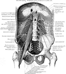
View of the posterior abdominal wall to show the muscles and the nerves of the lumbo sacral plexus.
Muscles of the Abdominal Wall

View of the posterior abdominal wall to show the muscles and the nerves of the lumbo sacral plexus.

Second layer of muscles of the back. Labels: 1, trapezius; 2, a portion of the ten dinous ellipse formed by the trapezius on both sides; 3, spine of scapula; 4, latissimus dorsi; 5, deltoid; 6, infra-spinatus and teres minor; 7, external oblique; 8, gluteus medius; 9, gluteus magnus; 10, levator scapulae; 11, rhomboideus minor; 12, rhomboideus major; 13, splenius capitis; 14, splenius colli; 15, portion of origin of latissimus dorsi; 16, serratus inferior posticus; 17, supra-spinatus; 18, infra-spinatus; 19, teres minor; 20, teres major; 21, long head of triceps extensor cubiti; 22, serratus major anticus; 23, internal oblique.

Muscles of the Back

Second layer of muscles of the back. Labels: 1, trapezius; 2, a portion of the ten dinous ellipse formed…

Muscles of the back. On the left side is exposed the first layer; on the right side, the second layer and part of the third.

Back Muscles

Muscles of the back. On the left side is exposed the first layer; on the right side, the second layer…

Deep layers of the muscles of the back.

Back Muscles

Deep layers of the muscles of the back.

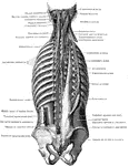
Deep muscles of the back.

Deep Back Muscles

Deep muscles of the back.

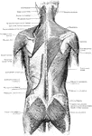
The superficial muscles of the back.

Superficial Muscles of the Back

The superficial muscles of the back.

Wooden box.

Box

Wooden box.

Boxes may be made of durable material such as wood or metal, or of corrugated fiberboard, paperboard, or other non durable materials.

Rectangular Box

Boxes may be made of durable material such as wood or metal, or of corrugated fiberboard, paperboard,…

The bud inserted.

The Bud, Inserted

The bud inserted.

A bud ready for insertion, and the T-shaped cut ready to receive it.

Bud, Ready for Insertion

A bud ready for insertion, and the T-shaped cut ready to receive it.

The budding completed

The Budding, Complete

The budding completed

A diagram showing six different parts of a grape vine. These parts include the shoot, cane, arm, branch, spur, and stem (trunk).

Ideal Vine for Cane Renewal

A diagram showing six different parts of a grape vine. These parts include the shoot, cane, arm, branch,…
"In makin Aeneas burn incense, Virgil follows the custom of his own time rather than historical verity." — Anthon, 1891

Ancient censer

"In makin Aeneas burn incense, Virgil follows the custom of his own time rather than historical verity."…

A side view of the chest and abdomen in respiration. Labels: 1, The cavity of the chest. 2, The cavity of the abdomen. 3, The line of direction for the diaphragm when relaxed in expiration. 4, The line of direction for the diaphragm when contracted in inspiration. 5, 6, The position of the front walls of the chest and abdomen in inspiration. 7, 8, The position of the front walls of the abdomen and chest in expiration.

A Side View of the Chest and Abdomen in Respiration

A side view of the chest and abdomen in respiration. Labels: 1, The cavity of the chest. 2, The cavity…

A diagram showing the separate abdominal and chest cavities.

Chest and Abdominal Cavity

A diagram showing the separate abdominal and chest cavities.

An illustration of two children, one standing and one resting on its knees, looking at an intricately carved box.

Children Looking at a Large Box

An illustration of two children, one standing and one resting on its knees, looking at an intricately…
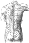
The distribution of cutaneous nerves n the back of the trunk. On the left side the distribution of the several nerves is represented, the letters indicating their nomenclature. ACR.; Posterior supraclavicular branches from cervical plexus; CIRC: Cutaneous branches of axillary nerve; E.C.: Lateral femoral cutaneous nerve, L.1: Lateral cutaneous branch of iliohypogastric nerve; S.1; Lateral branches of sacral nerves; T: T.1-12: Cutaneous branches of the posterior rami of the thoracic nerves. On the right side is a schematic representation of the areas supplied by the above nerves, the numeral and letter s indicate the spinal origins of the nerves distributed to each area.

Posterior View of the Cutaneous Nerves of Trunk

The distribution of cutaneous nerves n the back of the trunk. On the left side the distribution of the…

The distribution of the cutaneous nerves on the back of the trunk. On one side the distribution of the several nerves is represented, the letters indicating their nomenclature. Labels: G.O (C.2), great occipital; C.3, least occipital; T.1, et seq., posterior primary division of thoracic nerves; L.1, et seq., posterior primary divisions of first three lumbar nerves; S.1, et seq., posterior primary division of sacral nerves; Acr, acromial branches from cervical plexus; T.2-12, lateral branches of nerve; E.C, external cutaneous nerve; S.Sc, small sciatic nerve. One the other side a schematic representation is given of the areas supplied by the above nerves, the numeral indicating the spinal origin of the branches of distribution to each area.

Distribution of Cutaneous Nerves on the Back

The distribution of the cutaneous nerves on the back of the trunk. On one side the distribution of the…
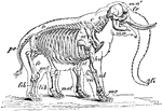
"Skeleton and Outline of African Elephant (Elephas or Loxodon africanus). fr, frontal; ma, mandible; ma', malar; fi, "finger" at end of trunk; C, cervical vertebrae; D, dorsal vertebrae; D, dorsal vertebrae; pe, pelvis; sc, scapula; st, sternum; hu, humerus; ul, ulna; ra, radius; mc, metacarpus; fe, femur; pat, patella; tib, tibia; fib, fibula; met, metatarsus." -Whitney, 1911

African Elephant Skeleton

"Skeleton and Outline of African Elephant (Elephas or Loxodon africanus). fr, frontal; ma, mandible;…
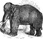
The elephant is the largest living land animal and is distinguished by its large ears and long trunk.

Indian Elephant

The elephant is the largest living land animal and is distinguished by its large ears and long trunk.

Man bending his trunk forward and backward.

Exercise

Man bending his trunk forward and backward.

Man bending his trunk sideways.

Exercise

Man bending his trunk sideways.

A barbed wire fence cutting in to a tree's trunk.

Fencepost, Cutting into a Tree

A barbed wire fence cutting in to a tree's trunk.

Ichthyophis glutinosus or Ceylon Caecilian is a species of amphibian in the Ichthyophiidae family. Pictured here is the ventral view of the head and trunk.

Head of Ichthyophis Glutinosus

Ichthyophis glutinosus or Ceylon Caecilian is a species of amphibian in the Ichthyophiidae family. Pictured…
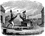
"A traveler's starting for a distant port, / The train is ready, and the time is short; / He gives a boy his trunk upon the way, / Bids him be quick, and not to stop and play, / But soon the boy pursues a butterfly, / Forgets his errand, and the time slips by."

If You Want a Thing Done, Go; If Not, Send

"A traveler's starting for a distant port, / The train is ready, and the time is short; / He gives a…
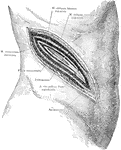
An incision in the right side above the kidney, showing a typical surgical approach to this organ, exposing the peritoneum and showing relations of abdominal muscles and fasciae.

Surgical Incision to the Kidney

An incision in the right side above the kidney, showing a typical surgical approach to this organ, exposing…
Trunks and luggage.

Luggage

Trunks and luggage.

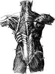
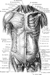
Superficial and deep muscles of the trunk. The sternocleidomastoid, pectoralis major, anterior portion of deltoid, the external oblique, aponeurosis of the internal oblique, tensor fasciae latae, sartorius, rectus femoris, iliopsoas, pectineus, adductor longus and the gracilis muscles of the left side have been cut away to expose the underlying muscles. Anterior View.

Anterior View of the Muscles of the Trunk

Superficial and deep muscles of the trunk. The sternocleidomastoid, pectoralis major, anterior portion…

Superficial and deep muscles of the trunk. The latissimus dorsi and trapezius on the right side have been cut away to expose the underlying muscles. Posterior View.

Posterior View of the Muscles of the Trunk

Superficial and deep muscles of the trunk. The latissimus dorsi and trapezius on the right side have…

"The Main Nerve Trunks of the Right Forearm, showing the Accompanying Radial and Ulnar Arteries. (Anterior view.)" — Blaisedell, 1904

Nerve trunks

"The Main Nerve Trunks of the Right Forearm, showing the Accompanying Radial and Ulnar Arteries. (Anterior…

The external oblique muscles

External Oblique Muscles

The external oblique muscles

The internal oblique muscles

Internal Oblique Muscles

The internal oblique muscles

The chief organs of the body from the side. Labels: a, arch of the aorta or main artery of the trunk; c, large intestine; d, diaphragm; e, throat; g, lung; k, kidney; l, spinal cord within the back bone; m, main artery divided to go to h legs; n, pancreas; o, gullet; s, spleen; tr, wind pipe; t, main vein of body; ap, appendix.

View of Organs from the Side

The chief organs of the body from the side. Labels: a, arch of the aorta or main artery of the trunk;…

"The <em>Palaeotherium magnum</em> was of the size of a horse, but thicker and more clumsy; its head was massive, and its legs and tail were short. It resembled a large tapir, but differed somewhat as to the teeth, and had one toe less on the fore-feet. Its height was probably from four to five feet; about equal, it is said, to that of the rhinoceros of Java. It was, no doubt, furnished with a short proboscis or trunk." &mdash; Goodrich, 1844

Palaeotherium magnum

"The Palaeotherium magnum was of the size of a horse, but thicker and more clumsy; its head…

"The <em>Palaeotherium minus</em> was smaller in size compared to the <em>Palaeotherium magnum</em>, probably not larger than a roe-buck, and of similar form to the tapir. It had light and slender limbs." &mdash; Goodrich, 1844

Palaeotherium minus

"The Palaeotherium minus was smaller in size compared to the Palaeotherium magnum,…
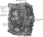
Section of human pancreas, showing pancreatic islands.

Section of Pancreas

Section of human pancreas, showing pancreatic islands.

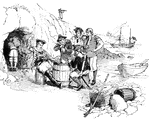
Pirates of the eighteenth century at their cave.

Pirates

Pirates of the eighteenth century at their cave.

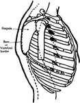
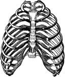
The trunk showing ribs.

Ribs

The trunk showing ribs.

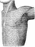
Lymphatics and lymphatic glands on the left side of the body and shoulder.

Lymphatics of the Shoulder

Lymphatics and lymphatic glands on the left side of the body and shoulder.

"The skeleton of the trunk and the limb arches seen from the front. C, clavicle; S, scapula; Oc, innominate bone attached to the side of the sacrum dorsally and meeting its fellow at the pubic symphysis in the ventral median line." &mdash;Martin, 1917

Skeletal Trunk

"The skeleton of the trunk and the limb arches seen from the front. C, clavicle; S, scapula; Oc, innominate…
Dissection of the spleen, liver, and kidneys from behind.

Spleen, Liver, and Kidneys

Dissection of the spleen, liver, and kidneys from behind.

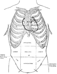
A diagram of the thoracic and abdominal regions. Labels: A, aortic valve; M, mitral valve; p, pulmonary valve; tricuspid valve.

Diagram of Thoracic and Abdominal Regions

A diagram of the thoracic and abdominal regions. Labels: A, aortic valve; M, mitral valve; p, pulmonary…
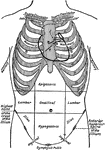
Diagram of the thoracic and abdominal regions. Labels: A, aortic valve; P, pulmonary valve; M, mitral valve; T, tricuspid valve.

Thoracic and Abdominal Regions

Diagram of the thoracic and abdominal regions. Labels: A, aortic valve; P, pulmonary valve; M, mitral…
The thorax consists of the dorsal vertebrae, the ribs, and the sternum.

Bony Walls of the Thorax

The thorax consists of the dorsal vertebrae, the ribs, and the sternum.

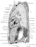
Lateral, sagittal section through the left thorax and upper portion of abdomen, viewed from the left. The plane of the section lies 4 cm to the left of the median plane.

Side View of the Thorax and Part of the Abdomen

Lateral, sagittal section through the left thorax and upper portion of abdomen, viewed from the left.…

The human torso. Labels: A, the heart; B, the lungs drawn aside to show the internal organs; C, the diaphragm; D, the liver; E, the gall cyst; F, the stomach; G, the small intestines; H, the transverse colon.

Torso

The human torso. Labels: A, the heart; B, the lungs drawn aside to show the internal organs; C, the…
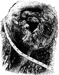
Inside of the broken limb, showing the decay that entered.

Inside the Tree Limb

Inside of the broken limb, showing the decay that entered.

A tree, which has been blown over due to a rotten trunk

A Tree, Blown Over

A tree, which has been blown over due to a rotten trunk

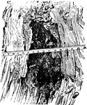
The decayed hole where a limb was removed. The wood-destroying fungi caused the tree to break.

A Hole in a Tree

The decayed hole where a limb was removed. The wood-destroying fungi caused the tree to break.