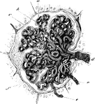
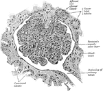
Malpighian Capsule
Malpighian capsule and tuft of capillaries, injected through the renal artery with colored gelatin.…
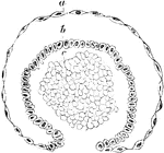
Development of Malpighian Capsule
Transverse section of a developing Malpighian capsule and tuft from a fetus at about the fourth month.…
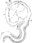
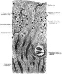
Epithelial Elements of a Malpighian Capsule
Epithelial elements of a Malpighian capsule and tuft, with the commencement of a urinary tubule showing…
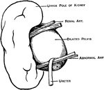
Abnormal Renal Artery
An abnormal renal artery causing kinking at the ureteropelvic junction, and hydronephrosis.
Side View of the Trunk
Sagittal section through the trunk, 6 cm to the right of the median plane, viewed from the right. Note…
Cross Section of the Trunk through Upper Kidney
Section through the upper pole of the left kidney at the level of the tip of the xiphoid process.
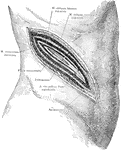
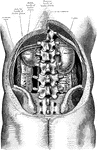
Surgical Incision to the Kidney
An incision in the right side above the kidney, showing a typical surgical approach to this organ, exposing…
Frontal Section Through Kidney
Frontal section through the right kidney and adjacent structures showing the renal fasciae and fatty…
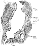
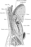
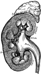
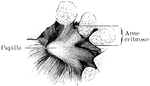
Sagittal Section Through Sinus of Kidney
Sagittal section through sinus of child's kidney, showing lower part of pelvis and commencement of ureter.
Longitudinal Section of Kidney of a Dog
Longitudinal section of injected kidney of dog, showing general arrangement of blood vessels of cortex…
Trunk Showing Organs of Digestion
Diagram of the relations of the large intestine and kidneys, from behind.
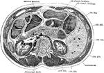
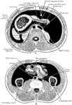
Transverse Section of the Trunk
Transverse section through the middle of the first lumbar vertebra, showing the relations of the pancreas.
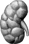
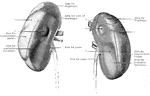
Anterior Surface of the Kidneys
The anterior surfaces of the kidneys, showing areas of contact of neighboring viscera.
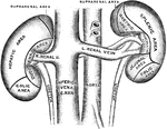
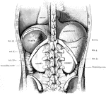
Posterior Surface of the Kidneys
The posterior surfaces of the kidneys, showing areas of relation to the parietes.
Section Through an Ape's Kidney
A section through the cortex of an ape's kidney. A Malpighian corpuscle, together with the beginning…
Section Through Kidney
Part of a section through the cortex of the kidney in the direction of the straight tubules.
Entrance of Ureter into the Bladder
Diagram showing the method of entrance of the ureter into the bladder.
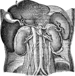
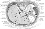
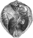
Abdominal Organs
Abdominal organs. Labels: 1, liver turned up; 2, gall bladder; 3, right kidney; 4, spleen; 5, left kidney.
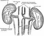
Horizontal Section Through Trunk
Diagram of horizontal section through upper part of 1st lumbar vertebra. The fine dots represent the…
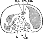
Transverse Section of Abdomen
Diagrammatic transverse section of abdomen, to show the peritoneum on transverse tracing. A, at level…
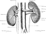
Kidneys from Behind
The kidneys viewed from behind. The dotted lines mark out the areas contact with the various muscle…
Kidneys from the Front
The kidneys and great vessels viewed from the front. The drawing was made before the removal of the…
Section Through Kidney
Longitudinal section through the kidney. The vessels and fat have been removed to give a view of the…
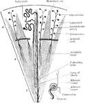
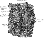
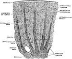
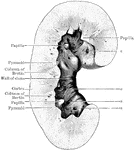
Structure of Kidney Lobe
Diagrammatic representation of the structure forming a kidney lobe. In the middle part of the figure…
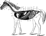
Digestive Apparatus of the Horse
The digestive apparatus of the horse. Labels: a, mouth; 2, pharynx; 3, esophagus; 4, diaphragm; 5, spleen;…
Liver and Diaphragm of a Horse
Posterior view of the liver and diaphragm in situ. Labels: a, left lobe; b, right lobe; c, quadrate…
Kidney of a Horse
Horizontal section of the right kidney. Labels: a, fibrous capsule detached; b, cortical layer; c, medullary…
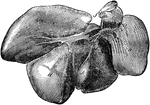
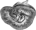
Liver of a Hog
Liver of a hog-posterior view. Labels: a, right external lobe; c, left external lobe; d, left internal…
Kidney of a Hog
Horizontal section of the kidney of a hog. Labels: a, cortical substance; b, medullary substance; c,…
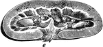
Earthworm Anatomy
The earthworms are also known as megadriles, in the families Tubificidae, Lumbriculidae, and Enchytraeidae.…
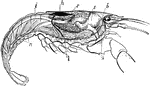
Crayfish
Crayfish, crawfish, or crawdads are freshwater crustaceans resembling small lobsters, to which they…
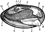
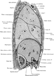
Mussel Anatomy
"Longitudinal Section through a Fresh-water Mussel. a, edge of mantle; b, foot, with position of ganglion…
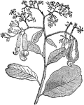
Cashew Branch
"Cashew Nut. Cashew, a tree common in the West Indies. Its fruit is called the cashew nut. The nut is…
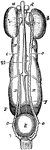
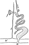
Female Uro-genital Organ
"Uro-genital organs of female embryo bird; from Owen, after Muller. a, testis; b, epididymis; c, sperm-duct…
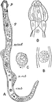
Dicyemennea Eledones Worm Found in Kidney of Octopus
A cross sectional view of the Dicyemennea Eledones, a parasite, found in Eledone Moschata, or musky…

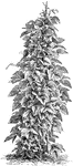
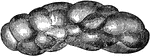
Runner or Climbing Kidney Bean
The common name of phaseolous multiflorus is runner beans or kidney beans. This plant grows tall and…
Anacardium Occidentale
The common name of Anacardium occidentale is cashew. The tree grows twenty to forty feet tall. The flowers…
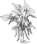
Kidney Bean
Common or kidney bean is the common name of Phaseolus vulgaris. In French it is known as haricot.
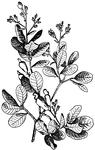
Cochlearia Danica
Cochlearia danica grows six to eight inches tall. The leaves are rounded and kidney shaped. The small…

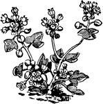
Ground Ivy
Ground Ivy (Glecoma hederacea) is a common European labiate hedgerow plant, with trailing stems, kidney-shaped,…
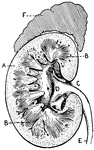
Section of Human Kidney
This illustration shows a section of a human kidney (A, Cortical substance; B, Pyramids; C, Hilum; D,…