The human humerus bone, the longest and largest bone of the upper leg. Labels: a, rounded head; gt, greater tuberosity; lt, lesser tuberosity; b, groove for attachment of biceps tendon.

The Human Humerus

The human humerus bone, the longest and largest bone of the upper leg. Labels: a, rounded head; gt,…

The Ulna and Radius. Labels: 1, radius; 2, ulna; o, olecranon process, on the anterior surface of which are seen the large (gs) and the small (ls) cavities for the reception of the lower end of the humerus and of the head of the radius, respectively; h, head of radius.

The Human Ulna and Radius

The Ulna and Radius. Labels: 1, radius; 2, ulna; o, olecranon process, on the anterior surface of which…
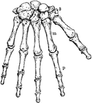
Bones of the Wrist and Hand. Labels: m, metacarpal bones; p, phalanges; 3, bones of wrist.

The Human Wrist and Hand Bones

Bones of the Wrist and Hand. Labels: m, metacarpal bones; p, phalanges; 3, bones of wrist.
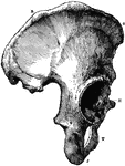
The Os Innominatum, or nameless bone, so called from bearing no resemblance to any known object, is a large irregular shaped bone, which, with its fellow of opposite side, forms the sides and front wall of the pelvic cavity. Labels: R, O, crest of ilium, just below O is seen the anterior superior spinous process; J, tuberosity of ischium; t, part of pubes, between J and T is seen the thyroid foramen: H, acetabulum, below H is seen end of ubic bone which, with its fellow of opposite side, forms the symphysis pubis.

Part of the Human Pelvic Bone

The Os Innominatum, or nameless bone, so called from bearing no resemblance to any known object, is…

The Femur (upper leg bone) is the longest, largest, and strongest bone in the skeleton. Labels: b, rounded head; n, neck; gtr, greater trochanter; ltr, lesser trochanter.

Human Femur Bone

The Femur (upper leg bone) is the longest, largest, and strongest bone in the skeleton. Labels: b, rounded…

Bones of the Ankle and Foot. Labels: m, metatarsal bones; p, phalanges; ca, os calcis, or heel bone.
Bones of the Ankle and Foot

Bones of the Ankle and Foot. Labels: m, metatarsal bones; p, phalanges; ca, os calcis, or heel bone.

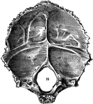
Occipital bone of the human skull, inner surface. It is situated at the back and base of the skull. Labels: 9,9, and 10,10, depression for reception of lobes of brain; 11, foramen magnum.

Occipital Bone of the Human Skull

Occipital bone of the human skull, inner surface. It is situated at the back and base of the skull.…
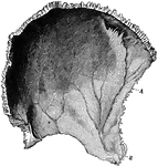
Parietal bone of the human skull, inner surface. The parietal bones form the greater part of the sides and roof of the skull. Labels: A, parietal depression; E, furrow for ramification of arteries.

Parietal Bone of the Human Skull

Parietal bone of the human skull, inner surface. The parietal bones form the greater part of the sides…

Frontal bone of the human skull, outer surface. The frontal bone forms the forehead, roof of the orbital cavities, and the nasal cavity. Labels: 1, frontal eminence; 7, roof of orbital cavitiy; 10, orbital arch.

Frontal Bone of the Human Skull

Frontal bone of the human skull, outer surface. The frontal bone forms the forehead, roof of the orbital…
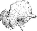
Temporal bone of the human skull. The temporal bones are situated at the sides and base of the skull. Labels: 1, squamous portion; 2, placed below external opening of auditory canal in petrous portion; 3, placed below mastoid portion; 4, placed below glenoid cavity for reception of condyle of lower jaw.

Temporal Bone of the Human Skull

Temporal bone of the human skull. The temporal bones are situated at the sides and base of the skull.…

Sphenoid bone, situated the anterior part of the base of the skull, articulating with all the other cranial bones, which it binds firmly and solidly together. Labels: a, greater wing; b, lesser wing.

Sphenoid Bone of the Human Skull

Sphenoid bone, situated the anterior part of the base of the skull, articulating with all the other…
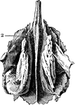
Ethmoid bone, posterior surface. The ethmoid bone is an exceedingly light, spongy bone, placed between the two orbits and at the root of the nose, contributing to form a part of each of these cavities. Label: 2, cribriform, or perforated plate.

Ethmoid Bone of the Human Skull

Ethmoid bone, posterior surface. The ethmoid bone is an exceedingly light, spongy bone, placed between…

Lachrymal Bone. The lachrymal are the smallest and most fragile bones fo the face. They are situated at the front part of the inner wall of the orbit.

Human Lachrymal Facial Bone

Lachrymal Bone. The lachrymal are the smallest and most fragile bones fo the face. They are situated…

Vomer bone, a single bone placed at the back part of the nasal cavity, and forms part of the septum of the nasal fossae.

Human Vomer Nasal Bone

Vomer bone, a single bone placed at the back part of the nasal cavity, and forms part of the septum…
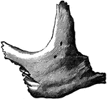
Malar (cheek) bone. The malar bones form the prominence of the cheek, and part of the outer wall and floor of the orbit.

Human Malar (Cheek) Bone

Malar (cheek) bone. The malar bones form the prominence of the cheek, and part of the outer wall and…
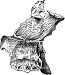
Palate bone. Palate bones form the back part of the roof of the mouth; part of the floor and outer wall of the nasal fossae, and a very small portion of the floor of the orbit.

Human Palate Bone

Palate bone. Palate bones form the back part of the roof of the mouth; part of the floor and outer wall…

Inferior turbinated bone, convex surface. The inferior turbinated bones are situated on the outer wall of each side of the nostril. Teach consists of a layer of thin, spongy bone, curled upon itself like a scroll.

Human Nostril Bone

Inferior turbinated bone, convex surface. The inferior turbinated bones are situated on the outer wall…
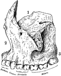
Superior maxillary bone. With it's fellow on the opposite side, it forms the whole of the upper jaw. Each bone assists in forming part of the floor of the orbit, the floor and outer wall of the nasal fossae, and the greater part of the roof of the mouth. Labels: 1, orbital surface; 2, facial surface; 3, alveolar process.

Human Maxillary (Upper Jaw) Bone

Superior maxillary bone. With it's fellow on the opposite side, it forms the whole of the upper jaw.…

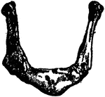
Inferior Maxillary Bone (lower jaw). It is the largest and strongest bone in the face and serves for the reception of the lower teeth.

Human Maxillary (Upper Jaw) Bone

Inferior Maxillary Bone (lower jaw). It is the largest and strongest bone in the face and serves for…
The hyoid, os hyoides, or tongue bone, is an isolated, U-shaped bone lying in front of the throat, just above the Adam's apple; it supports the tongue, and give attachment to some of its numerous muscles.

Human Hyoid Bone

The hyoid, os hyoides, or tongue bone, is an isolated, U-shaped bone lying in front of the throat, just…
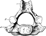
A cervical vertebra of the spine, inferior surface. Labels: 1, spinous process, slightly bifid; 4, transverse process; 5, articular process, inferior surface. Below the arch, or hollow portion, is seen the solid portion, or body.

Human Cervical Vertebra Bone

A cervical vertebra of the spine, inferior surface. Labels: 1, spinous process, slightly bifid; 4, transverse…

Side view of spinal column, without sacrum and coccyx. Labels: 1 to 7, cervical vertebrae; 8 to 19, dorsal vertebrae; 20 to 24, lumbar vertebrae; A, A, spinous processes; C, D, transverse processes; E, intervertebral aperture or foramen; a, atlas; 2, axis.

Human Spinal Column

Side view of spinal column, without sacrum and coccyx. Labels: 1 to 7, cervical vertebrae; 8 to 19,…

Thorax. The thorax, or chest, is an elongated conical-shaped cage, formed by the sternum and costal cartilages in front, the 12 ribs on each side, and the bodies of the 12 dorsal vertebrae behind. It contains and protects the principal organs of respiration and circulation. Labels: 1 to 12, ribs; d, d, costal cartilages; e, upper end of sternum; b, middle portion of sternum; 1 a, first dorsal vertebra; 12 a, twelfth dorsal vertebra; 7 a, seventh cervical vertebra; 1 to 7, true ribs; 8 to 12, false ribs; 11, 12, floating ribs. 10th rib is defective; it should be attached to the costal cartilage.

Human Thorax (Chest)

Thorax. The thorax, or chest, is an elongated conical-shaped cage, formed by the sternum and costal…

Sternum, front and side view. The sternum, or breast bone, is a flat narrow bone, situated in the median line in the front of the chest, and consisting, in the adult, of 3 portions.

Human Sternum Bone

Sternum, front and side view. The sternum, or breast bone, is a flat narrow bone, situated in the median…
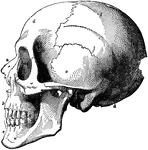
The skull. Labels: a, nasal bone; b, superior maxillary; c, inferior maxillary; d, occipital; e, temporal; f, parietal; g, frontal bone.

Human Skull

The skull. Labels: a, nasal bone; b, superior maxillary; c, inferior maxillary; d, occipital; e, temporal;…
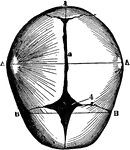
The skull at birth, superior suerface. The cranial bones of the infant at birth are not fullyformed and their edges are separated by membranous intervals. Labels: 1, posterior fontanelle; 2, sagital suture; 4, anterior fontanelle; A, A, bi-parietal diameter; B, B, bi-temporal diamter.

Human Skull at Birth

The skull at birth, superior suerface. The cranial bones of the infant at birth are not fullyformed…

Male pelvis (top) and female pelvis (bottom). The pelvis is stronger and more massively constructed than either the cranial or thoracic cavity. It is composed of 4 bones, the ossa innominata, forming sides and front, and the sacrum and coccyx, completing it behind. The female pelvis differs from that of the male in making it better adapted for giving birth. The most notable differences are that the female pelvis is wider in every direction, giving more room for the child to pass and shallower, which lessens the distance through which the child has to be propelled; and lastly, the bones are thinner and smoother.

Human Pelvis, Male and Female

Male pelvis (top) and female pelvis (bottom). The pelvis is stronger and more massively constructed…

A mixed articulation (slightly movable). In this form, the bony surfaces are usually joined together by broad, flattened disks of fibro-cartilage, as in the articulations between the bodies of the vertebrae. Labels: a, b, disk of fibro-cartilage; c, articular cartilage; d, bone.

Human Joint, Mixed Articulation

A mixed articulation (slightly movable). In this form, the bony surfaces are usually joined together…
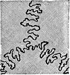
A toothed, or dentated suture. This is one type of immovable articulation. It is found in the union of the cranial bones of the skull, such as the suture between the frontal and parietal bones.

Human Joint, Dentated Suture

A toothed, or dentated suture. This is one type of immovable articulation. It is found in the union…
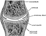
A simple complete joint, one type of movable articulation. The synovial membrane is represented by dotted lines. They have a secreting membrane placed between their opposing surfaces, which keeps them well lubricated and capable of free movement one upon the other.

A Simple Complete Joint

A simple complete joint, one type of movable articulation. The synovial membrane is represented by dotted…
The bony and cartilaginous skeleton.

Human Skeleton, Showing Bony and Cartilage Tissue

The bony and cartilaginous skeleton.

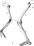
The skeleton of the arm and leg. Labels: H, the humerus; Cd, its articular head which fits into the glenoid fossa of the scapula; U, the ulna; R, the radius; O, the olecranon; Fe, the femur; P, the patella; Fi, the fibula; T, the tibia.

Arm and Leg Skeleton

The skeleton of the arm and leg. Labels: H, the humerus; Cd, its articular head which fits into the…

The skeleton of the trunk and the limb arches seen from the front. Labels: c, clavicle; S, scapula; Oc, innominate bone attached to the side of the sacrum dorsally and meeting its fellow at the pubic symphysis in the ventral median line.

Skeleton of Trunk

The skeleton of the trunk and the limb arches seen from the front. Labels: c, clavicle; S, scapula;…

Demonstration of the movement of a pivot joint. Labels: A, arm in supination (palm uppermost); B, arm in pronation (back of hand upward). H, humerus; R, radius; U, ulna.

Arm Bones

Demonstration of the movement of a pivot joint. Labels: A, arm in supination (palm uppermost); B, arm…
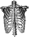
The skeleton of the thorax. Labels: a, g, vertebral column; b, first rib; c, clavicle; d, third rib; i, glenoid fossa.

Thorax

The skeleton of the thorax. Labels: a, g, vertebral column; b, first rib; c, clavicle; d, third rib;…
Front view of the body and outline of skeleton and covering.

Front View of the Body

Front view of the body and outline of skeleton and covering.

Back view of the body and outline of skeleton and covering.

Back View of the Body

Back view of the body and outline of skeleton and covering.

The human skeleton.

Human Skeleton

The human skeleton.

The bony and cartilaginous skeleton.

The Bony and Cartilaginous Skeleton

The bony and cartilaginous skeleton.

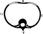
Diagrammatic representation of a segment of the axial skeleton V, a vertebra; C, Cv, ribs articulating above with the body and transverse process of the vertebra; S, the breastbone. The lighter-shaded part between S and C is the costal cartilage.

Segment of the Axial Skeleton

Diagrammatic representation of a segment of the axial skeleton V, a vertebra; C, Cv, ribs articulating…
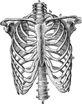
The human skeleton.

The Human Skeleton

The human skeleton.

The skeleton of the thorax. Labels: a, g, vertebral column; b, first rib; c, clavicle; e, seventh rib; i, glenoid fossa.

Thorax Skeleton

The skeleton of the thorax. Labels: a, g, vertebral column; b, first rib; c, clavicle; e, seventh rib;…

The human skeleton.

Human Skeleton

The human skeleton.

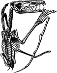
The skeleton of a pterodactyl, the prehistoric winged dinosaur.

Pterodactyl Skeleton

The skeleton of a pterodactyl, the prehistoric winged dinosaur.

Front view of the human skeleton.

Skeleton, Front View

Front view of the human skeleton.

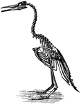
The Hesperornis regalis was an ancient bird of the Cretaceous Period.

Hesperornis Regalis

The Hesperornis regalis was an ancient bird of the Cretaceous Period.

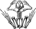
The skeleton of a frog.

Frog Skeleton

The skeleton of a frog.

"Skull of Boa constrictor. a, quadrate bone; b, b, halves of lower jaw." -Cooper, 1887

Boa Constrictor Skull

"Skull of Boa constrictor. a, quadrate bone; b, b, halves of lower jaw." -Cooper, 1887

The leaves of the skeleton-leaved geranium, Pelargonium radula.

Skeleton-Leaved Germanium

The leaves of the skeleton-leaved geranium, Pelargonium radula.

"Restored skeleton of Palaeospondylus gunni. d.c., Cirri of dorsal margin; l.c., long lateral cirri; v.c., cirri of ventral margin; n., nasal ring; t.p., anterior trabeculo-palatine part of cranium; b., anterior depression or fenestra; c., posterior depression or fenestra; a., lobe divided off from anterior part; p.a., posterior or parachordal part of cranium; x., post-occipital plates." -Thomson, 1916

Palaeospondylus Gunni

"Restored skeleton of Palaeospondylus gunni. d.c., Cirri of dorsal margin; l.c., long lateral cirri;…
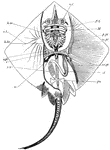
Skeleton of skate. nc, nasal capsules; pq, palato-pterygo-quadrate cartilage; M, Meckel's cartilage; hm, hyo-mandibular; h.br., fifth hypobranchial; v.pl., vertebral plate; st, sensory tubes; sc, scapular region; c, coracoid region; p.pt., anterior basal cartilage or pro-pterygium; m.pt., meso pterygium; mt.pt., meta-pterygium; pu., pubic bar; st, stomach; sv, spiral valve of intestine. -Thomson, 1916

Skate Skeleton

Skeleton of skate. nc, nasal capsules; pq, palato-pterygo-quadrate cartilage; M, Meckel's cartilage;…
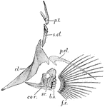
"Pectoral girdle and fin of cod. fr., Fin-rays; b.o., brachial ossicles; cor., coracoid; sc., scapula; cl., clavicle; p.cl., post-clavicle; s.cl., supra-clavicle; p.t., post-temporal." -Thomson, 1916

Cod Fin

"Pectoral girdle and fin of cod. fr., Fin-rays; b.o., brachial ossicles; cor., coracoid; sc., scapula;…

"Skeleton of Ceratodus fin. a., Central axis; r., radials; h., basal piece." -Thomson, 1916

Queensland Lungfish Fin

"Skeleton of Ceratodus fin. a., Central axis; r., radials; h., basal piece." -Thomson, 1916

"Vertebral column and pelvic girdle of bull-frog. t.p., Transvers processes of sacral vertebra; Il., ilium; U., urostyle; Fe., femur; Isch., ischiac region." -Thomson, 1916

Bullfrog Vertebrae

"Vertebral column and pelvic girdle of bull-frog. t.p., Transvers processes of sacral vertebra; Il.,…
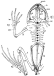
"Skeleton of frog. The half of the pectoral girdle, and fore- and hind-limb of the right side are not shown. pmx., premaxilla; m.x., maxilla; n., nasal; sph., sphenethmoid; p.f., parieto-frontal; P.O., pro-otic; pt., pterygoid; q.j., quadrato-jugal; sq., squamosal; Q., quadrate; c., columella auris; A., atlas; t.p., transverse process; S.V., sacral vertebra; U., urostyle; S.sc., supra scapula; H., humerus; R.U., radio ulna; Cp., carpals; Mc., metacarpals; Il., ilium; Is., ischium; F., femur; T.F., tibio-fibula; Ca., calcaneum; As., astragalus; C., calcar; Mt., metatarsals." -Thomson, 1916

Frog Skeleton

"Skeleton of frog. The half of the pectoral girdle, and fore- and hind-limb of the right side are not…
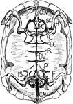
"Internal view of tortoise skeleton. H., humerus; SC., scapula running dorsally; PC., precoracoid; C., coracoid; EC., epicoracoid cartilage; P., pubis; IL., ilium running dorsally to sacral vertebrae; IS., ischium; DV., dorsal vertebrae fused in carapace; R., head of a rib; CEV., cervical vertebrae free; CAV., caudal vertebrae free." -Thomson, 1916

Tortoise Skeleton

"Internal view of tortoise skeleton. H., humerus; SC., scapula running dorsally; PC., precoracoid; C.,…
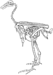
"Entire skeleton of condor, showing the relative positions of the chief bones." -Thomson, 1916

Condor Skeleton

"Entire skeleton of condor, showing the relative positions of the chief bones." -Thomson, 1916

"Hesperornis. ST., Sternum; CO., coracoid; CL., clavicle; H., rudimentary humerus; SC., scapula; P., pectineal pubic process; IL., ilim; IS., ischium; P.P., post-pubis; C.T., crest of tibia; F. fibula; T.T., base tibio-tarsus; T.M.T., tarso-metatarsus." -Thomson, 1916

Hesperornis Skeleton

"Hesperornis. ST., Sternum; CO., coracoid; CL., clavicle; H., rudimentary humerus; SC., scapula; P.,…

"Skeleton of male gorilla. cl., Clavicle; sc., tip of scapula; S., praesternum; H., humerus; r., radius; u., ulna; Il., ilium; C., coccyx; P., pubis; Is., ischium; F., femur; t., tibia; f., fibula." -Thomson, 1916

Gorilla Skeleton

"Skeleton of male gorilla. cl., Clavicle; sc., tip of scapula; S., praesternum; H., humerus; r., radius;…
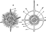
"Actinomma, a radiolarian with a shell and no mouth. A, whole animal with a portion of two spheres of shell removed. B, section, showing relation of protoplasm to the skeleton. c., central capsule; n, nucleus; p, protoplasm; o, openings through which the pseudopodia extend." -Galloway, 1915

Actinomma

"Actinomma, a radiolarian with a shell and no mouth. A, whole animal with a portion of two spheres of…