A diagram of the relative position of the bone, cartilage, and synovial membrane. Labels: 1,The extremities of two bones to form a joint. 2, The cartilage that covers the end of the bone. 3, The synovial membrane, which covers the cartilage of both bones and is then doubled back from one to the other, represented by the dotted lines.

Position of the Bone, Cartilage, and Synovial Membranes

A diagram of the relative position of the bone, cartilage, and synovial membrane. Labels: 1,The extremities…

"Cartilage or gristle is fibrous tissue glued together by a substance containing chondrine." — Richardson, 1906

Cartilage

"Cartilage or gristle is fibrous tissue glued together by a substance containing chondrine." —…
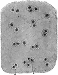
Portions of cartilage seen in section.

Cartilage Portion in Section

Portions of cartilage seen in section.

A thin slice of cartilage highly magnified, showing the cartilage cells (a,b) scattered through an almost structureless material.

Cartilage Tissue

A thin slice of cartilage highly magnified, showing the cartilage cells (a,b) scattered through an almost…

Fibrous cartilage connective tissue from the symphysis pubis, magnified. Cartilage is a structure without blood vessels and nerves, containing cells lying in a dense substance, which on boiling becomes jelly-like, resembling glue. It is mainly found in the joints, but also in the ear, nose, and air passages of the chest.

Cartilage Tissue

Fibrous cartilage connective tissue from the symphysis pubis, magnified. Cartilage is a structure without…

"Showing (1) cartilage with martrix and cells; (2) cartilage with matrix containing cells and white fibers." — Blaisedell, 1904

Longitudinal section of cartilage

"Showing (1) cartilage with martrix and cells; (2) cartilage with matrix containing cells and white…

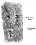
Cartilage is made up of string-like fiber and few cells.

White Fiber Cartilage

Cartilage is made up of string-like fiber and few cells.

"Membranous cartilage skull of <em>Cyclostoma</em>."&mdash;Finley, 1917

Skull of Cyclostoma

"Membranous cartilage skull of Cyclostoma."—Finley, 1917

"Median longitudinal section of anterior region of Myxine. B., Barbule; N., nasal aperture; NT., nasal tube with rings of cartilage; NC., nasal capsule; BR., brain; SC., spinal cord; N., notochord; G., gut; T., cartilage of "tongue"; MU., muscle; NTT., posterior part of nasal sac; TP., a tooth plate; MT., median tooth on roof of mouth (M.)." -Thomson, 1916

Hagfish Anterior

"Median longitudinal section of anterior region of Myxine. B., Barbule; N., nasal aperture; NT., nasal…
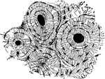
The humerus bisected lengthwise. Labels: a, marrow-cavity; b, hard bone; c, spongy bone; d, articular cartilage.

Bisection of the Humerus

The humerus bisected lengthwise. Labels: a, marrow-cavity; b, hard bone; c, spongy bone; d, articular…

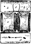
A joint between two bones (a and b). The ends of all bones are tipped with cartilage so that they may move easily upon each other. And besides this, the ends are lined with a very fine membrane, so arranged as to make a closed sac (c).

Joint

A joint between two bones (a and b). The ends of all bones are tipped with cartilage so that they may…
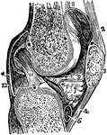
A vertical section of the knee joint. Labels: femur; 3, patella; 2, 4, ligaments of the patella; 5, tibia; 6, cartilage of the tibia; 12, cartilage of the membrane.

Vertical Section of the Knee Joint

A vertical section of the knee joint. Labels: femur; 3, patella; 2, 4, ligaments of the patella; 5,…
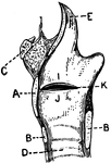
This illustration shows a vertical section of the larynx and its many parts (A. Thyroid Cartilage; B. Cricoid Cartilage; C. hyoid bone; D. Rings of trachea; E. Epiglottis; F. Arytenoid Cartilage; G. Thyro-arytenoid muscle; H. Adam's Apple; I. False Vocal Cords; J. True Vocal Cords; K. Ventricle; L. Rima Glottis).

Vertical Section of Larynx

This illustration shows a vertical section of the larynx and its many parts (A. Thyroid Cartilage; B.…

Arytenoid cartilages of the larynx. These are pitcher-like cartilages that are 2 in number, pyramidal-shaped, and situated on the upper margin of the cricoid cartilage posteriorly, closing in the back of the larynx.

Arytenoid Cartilages of the Larynx

Arytenoid cartilages of the larynx. These are pitcher-like cartilages that are 2 in number, pyramidal-shaped,…

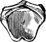
Cricoid cartilage of the larynx, situated below the thyroid.

Cricoid Cartilage of the Larynx

Cricoid cartilage of the larynx, situated below the thyroid.

"Cartilages and Ligaments of the Larynx. (Front view.) <em>A</em>, hyoid bone; <em>B</em>, membrane attached to hyoid bone and the shield-shaped cartilage below (thyroid); edges of this shield-shaped cartilage unite at <em>C</em> (Adam's apple is the V-shaped groove on a line with <em>B</em> and <em>C</em>; <em>D</em>, membrane between the shield-shaped cartilage and the signet-ring cartilage below; <em>E</em> cricoid, or signet-ring, cartilage; <em>F</em>, upper ring of the windpipe." &mdash; Blaisedell, 1904

Front view of the larynx

"Cartilages and Ligaments of the Larynx. (Front view.) A, hyoid bone; B, membrane…

"Cartilages and Ligaments of the Larynx. (Front view.) <em>A</em>, epiglottis; <em>B</em>, thyroid cartilage; <em>C</em>, ladle-shaped cartilage; <em>E</em>, cricoid cartilage; <em>F</em>, upper ring of the windpipe." &mdash; Blaisedell, 1904

Posterior view of the larynx

"Cartilages and Ligaments of the Larynx. (Front view.) A, epiglottis; B, thyroid cartilage;…
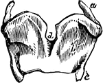
Thyroid cartilages of the larynx.

Thyroid Cartilages of the Larynx

Thyroid cartilages of the larynx.

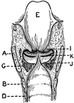
This illustration shows a longitudinal section of  the larynx as seen from behind (A. Thyroid Cartilage; B. Cricoid Cartilage; C. hyoid bone; D. Rings of trachea; E. Epiglottis; F. Arytenoid Cartilage; G. Thyro-arytenoid muscle; H. Adam's Apple; I. False Vocal Cords; J. True Vocal Cords; K. Ventricle; L. Rima Glottis).

Longitudinal Section of Larynx Seen from Behind

This illustration shows a longitudinal section of the larynx as seen from behind (A. Thyroid Cartilage;…
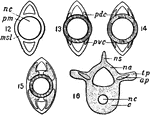
"Development of notochord : msl, mesoblastic skeletogenous layer ; pdc, paired dorsal cartilage; pvc, paired ventral cartilage; ns, neautral spine; na, neutral arch; tp, transverse process; articular process; c, centrum."&mdash;Finley, 1917

Development of the notochord

"Development of notochord : msl, mesoblastic skeletogenous layer ; pdc, paired dorsal cartilage; pvc,…

"The bones of fishes are of a less dense and compact nature than in the higher order of animals; in some, indeed, they are wholly cartilaginous. The skeleton may in general be divided into four chief parts - the Vertical Column, the Head, the Repiratory Apparatus, and the Limbs" &mdash; Goodrich, 1859

Perch skeleton

"The bones of fishes are of a less dense and compact nature than in the higher order of animals; in…

"Cartilaginous Cranium of the Pike (Esox lucius), with its intrinsic ossifications. A, top view; 3, small ossifications in the rostrum; N, N, nasal fossae; I Or, interorbital septum; Eth, ethmoid; Prf, Ptf, prefrontal and postfrontal; EpO, epiotic; OpO, opisthotic; Pt, pteriotic; EO, exoccipital." -Whitney, 1911

Pike Cranium

"Cartilaginous Cranium of the Pike (Esox lucius), with its intrinsic ossifications. A, top view; 3,…

"Cartilaginous Cranium of the Pike (Esox lucius), with its intrinsic ossifications. B, side view: V, VII, exits of trigeminal and of pneumogastric nerves; 3, small ossifications in the rostrum; I Or, interorbital septum; Eth, ethmoid; Prf, Ptf, prefrontal and postfrontal; PrO, pro&ouml;tic; EpO, epiotic; OpO, opisthotic; Pt, pteriotic; EO, exoccipital; BO, basioccipital; BS, basisphenoid; AS, alisphenoid." -Whitney, 1911

Pike Cranium

"Cartilaginous Cranium of the Pike (Esox lucius), with its intrinsic ossifications. B, side view: V,…
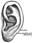
"The outer ear consists of a plate of gristle, shaped somewhat like a shell, known as the pinna, or auricle, and of a tube about an inch long, called the auditory canal." &mdash; Blaisedell, 1904

Pinna

"The outer ear consists of a plate of gristle, shaped somewhat like a shell, known as the pinna, or…
Inner surface of cartilage of pinna.

Inner Surface of Pinna Cartilage

Inner surface of cartilage of pinna.

Outer surface of cartilage of pinna.

Outer Surface of Pinna Cartilage

Outer surface of cartilage of pinna.

"Lateral view of caudal vertebra of Basking Shark (Selache mazima). a, centrum; b, neurapophysis; c, intercrural cartilage; d, hemapophysis; e, spinal canal; f, intervertebal cavity; g, central canal for persistent portion of notochord; h, hemal canals for blood-vessels." &mdash; Encyclopedia Britannica, 1893

Shark Vertebra

"Lateral view of caudal vertebra of Basking Shark (Selache mazima). a, centrum; b, neurapophysis; c,…
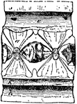
"Longitudinal section of caudal vertebra of Basking Shark (Selache mazima). a, centrum; b, neurapophysis; c, intercrural cartilage; d, hemapophysis; e, spinal canal; f, intervertebal cavity; g, central canal for persistent portion of notochord; h, hemal canals for blood-vessels." &mdash; Encyclopedia Britannica, 1893

Shark Vertebra

"Longitudinal section of caudal vertebra of Basking Shark (Selache mazima). a, centrum; b, neurapophysis;…

"Transverse section of caudal vertebra of Basking Shark (Selache mazima). a, centrum; b, neurapophysis; c, intercrural cartilage; d, hemapophysis; e, spinal canal; f, intervertebal cavity; g, central canal for persistent portion of notochord; h, hemal canals for blood-vessels." &mdash; Encyclopedia Britannica, 1893

Shark Vertebra

"Transverse section of caudal vertebra of Basking Shark (Selache mazima). a, centrum; b, neurapophysis;…
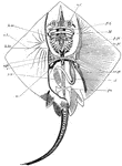
Skeleton of skate. nc, nasal capsules; pq, palato-pterygo-quadrate cartilage; M, Meckel's cartilage; hm, hyo-mandibular; h.br., fifth hypobranchial; v.pl., vertebral plate; st, sensory tubes; sc, scapular region; c, coracoid region; p.pt., anterior basal cartilage or pro-pterygium; m.pt., meso pterygium; mt.pt., meta-pterygium; pu., pubic bar; st, stomach; sv, spiral valve of intestine. -Thomson, 1916

Skate Skeleton

Skeleton of skate. nc, nasal capsules; pq, palato-pterygo-quadrate cartilage; M, Meckel's cartilage;…

"Under surface of skull and arches of skate. l.1, First labial cartilage; R., rostrum; tr., trabecular region; n.c., nasal capsule; a.o., antorbital cartilage; p.pt.q., palato-pterygo-quadrae; M.c., Meckel's cartilage; h.m., hyo-mandibular; h.br.1-5, hypobranchials; c.br.5, fifth ceratobranchial; c.h., cerato-hyal; l.2-4, labial cartilages." -Thomson, 1916

Skate Skull

"Under surface of skull and arches of skate. l.1, First labial cartilage; R., rostrum; tr., trabecular…

"Side view of skate's skull. l1., First labial cartilage; n.c., nasal capsule; a.o., antorbital; p.pt.q., palato-pterygo-quadrate; M.c., Meckel's cartilage; h.m., hyo-mandibular; e.h., epi-hyal; c.h., cerato-hyal; h.h., hypo-hyal; h.br.1-5, hypobranchials; c.br., ceratobranchial; e.br., epibranchial; p.br.1., first prebranchial; i.h., inter-hyal; m.pt., meta-pterygoid; 2, 5, 7, foramina of exit of the corresponding nerves." -Thomson, 1916

Skate Skull

"Side view of skate's skull. l1., First labial cartilage; n.c., nasal capsule; a.o., antorbital; p.pt.q.,…
The bony and cartilaginous skeleton.

Human Skeleton, Showing Bony and Cartilage Tissue

The bony and cartilaginous skeleton.

The bony and cartilaginous skeleton.

The Bony and Cartilaginous Skeleton

The bony and cartilaginous skeleton.

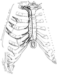
The anterior (ventral) surface of the sternum and the costal cartilages.

Sternum

The anterior (ventral) surface of the sternum and the costal cartilages.

The posterior (dorsal) surface of the sternum.

Sternum

The posterior (dorsal) surface of the sternum.

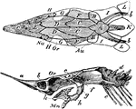
"Top and side views. The cartilaginous cranium shaded, is supposed to be seen through the unshded canial bones."-Whitney, 1902

Sturgeon Skull

"Top and side views. The cartilaginous cranium shaded, is supposed to be seen through the unshded canial…
A side view of the thyroid and cricoid cartilages.

Thyroid

A side view of the thyroid and cricoid cartilages.

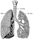
"The <em>trachea</em> has in its walls stiff rings of cartilage that hold it open so that the air can pass freely through it to and from the lungs. At its base the trachea divides and sends a great branch to each lung." &mdash; Ritchie, 1918

Trachea and lungs

"The trachea has in its walls stiff rings of cartilage that hold it open so that the air can…
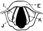
This illustration shows the vocal cords as seen from above during phonation (A. Thyroid Cartilage; B. Cricoid Cartilage; C. hyoid bone; D. Rings of trachea; E. Epiglottis; F. Arytenoid Cartilage; G. Thyro-arytenoid muscle; H. Adam's Apple; I. False Vocal Cords; J. True Vocal Cords; K. Ventricle; L. Rima Glottis).

Vocal Cords Seen from above During Phonation

This illustration shows the vocal cords as seen from above during phonation (A. Thyroid Cartilage; B.…
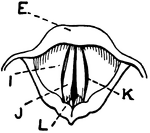
This illustration shows the vocal cords, seen from above during quiet breathing (A. Thyroid Cartilage; B. Cricoid Cartilage; C. hyoid bone; D. Rings of trachea; E. Epiglottis; F. Arytenoid Cartilage; G. Thyro-arytenoid muscle; H. Adam's Apple; I. False Vocal Cords; J. True Vocal Cords; K. Ventricle; L. Rima Glottis).

Vocal Cords Seen from above During Quiet Breathing

This illustration shows the vocal cords, seen from above during quiet breathing (A. Thyroid Cartilage;…