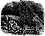
'Tis All for the Best
The picture describes a story about a traveler who encounters a terrible storm and because he cannot…
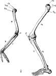
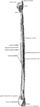
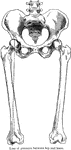
Arm and Leg Skeleton
The skeleton of the arm and leg. Labels: H, the humerus; Cd, its articular head which fits into the…
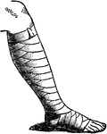
Roller Bandage
"Roller Bandage. Bandage, a surgical wrapper of some kind applied to a limb or other portion of the…
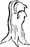
Leg of Bear
This illustration shows the plantigrade leg of a bear. Plantigrade means that the animal walks flat…
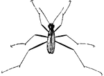
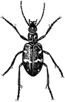
Wood Tiger Beetle
A group of beetles known for being preditors. They have bulging eyes and slender legs.
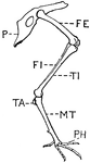
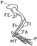
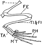
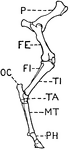
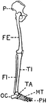
Leg of Bird
This illustration shows the leg of a bird. P. Pelvis, FE. Femur, TI. Tibia, FI. Fibula, TA. Tarsus,…
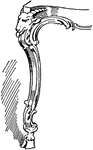
Leg of Candelabrum Base
This leg of candelabrum base is an antique design that is found in Paestum, Italy. In this design the…
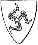
Charge
Human legs are not unfrequently born as charges in Heraldry, sometimes naked, sometimes booted, and…
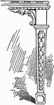
Chippendale Table Leg 1
A table leg designed by Thomas Chippendale. This table leg has a "cabriole" style.
Chippendale Table Leg 2
A table leg designed by Thomas Chippendale. The table leg has a taper leg style.
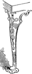
Chippendale Table Leg 3
A table leg designed by Thomas Chippendale. This table leg has a "cabriole" leg style.
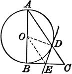
Circle With a Right Triangle
Illustration where one leg of a right triangle is the diameter of a circle. The tangent at the point…
Leg of Crocodile
This illustration shows the leg of a crocodile. P. Pelvis, FE. Femur, TI. Tibia, FI. Fibula, TA. Tarsus,…
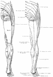
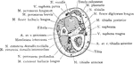
Cutaneous Nerves of the Back Legs
Distribution of cutaneous nerves on the back of the lower legs. On the one side the distribution of…
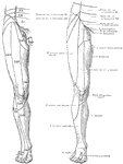
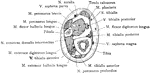
Cutaneous Nerves on the Back of the Legs
Distribution of cutaneous nerves on the back of the right lower extremity. The figure at the right shows…
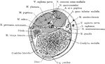
Cutaneous Nerves on the Front of the Legs
Distribution of cutaneous nerves on the front of the right lower extremity. The figure at the right…
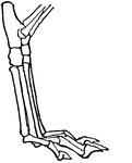
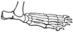
Leg of Dog
This illustration shows the leg of a dog. This leg is digitigrade. Animals with digitigrade legs walk…
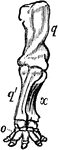
Anterior Extremity of Elephant
"Shows how the bones of the arm (q), forearm (q'x), and foot (o), are twisted to form an osseous screw."—Pettigrew,…
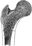
Frontal Section Through Upper End of Femur
Frontal section through upper end of femur, showing arrangement of pressure and tension lamellae.
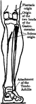
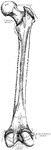
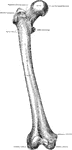
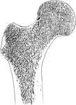
Human Femur Bone
The Femur (upper leg bone) is the longest, largest, and strongest bone in the skeleton. Labels: b, rounded…
Longitudinal Section of the Femur
The longitudinal section of the extremity of the femur, exhibiting the arrangement of the spongy substance.…
Cross Section Five Inches Above the Lower End of the Fibula
Section five inches above the lower end of the fibula.
Cross Section Four Inches Above the Lower End of the Fibula
Section four inches above the lower end of the fibula.
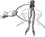
Frog Experiment
"Galvani found that whenever the nerves of a frog's leg were touched by one metal and the muscles by…
Leg of Frog
This illustration shows the leg of a frog. P. Pelvis, FE. Femur, TI. Tibia, FI. Fibula, TA. Tarsus,…
Gambe
"GAMBE. An obsolete French word, signifying a leg, and is still used in Heraldry, for the leg of a lion…
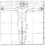
Proportions of human figure
"The proportions of the human figure. As handed down to us by Vitruvius and described by Joseph Bonomi."…
Human Leg (Front View)
This illustration shows a front view of a human leg. P. Pelvis, FE. Femur, TI. Tibia, FI. Fibula, TA.…
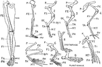
Human Leg (Front View), and Comparative Diagrams showing Modifications of the Leg
This illustration shows a human leg (front view), and comparative diagrams showing modifications of…
Human Leg (Side View)
This illustration shows a side view of a human leg. P. Pelvis, FE. Femur, TI. Tibia, FI. Fibula, TA.…
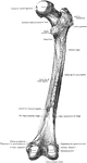
The Human Humerus
The human humerus bone, the longest and largest bone of the upper leg. Labels: a, rounded head; gt,…
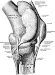
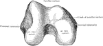
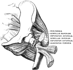
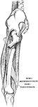
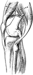
Knee Joint from Lateral Surface
Right knee joint from the lateral surface. The joint cavity and several bursae have been injected with…
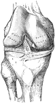
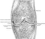
Frontal Section Through Knee Joint
Frontal section through middle of right knee joint. Seen from behind.
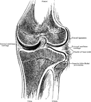
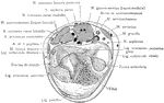
Frontal Section Through Knee Joint
Frontal section through knee joint, showing articulating surfaces and epiphyseal lines.