Clipart tagged: ‘maxilla’
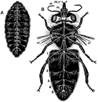
Bee Nervous System
"Nervous system of bee. A, of larva. B, of adult. a., Antenna; mx., maxilla; m., mandible; w., origin…
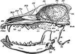
Cape Jumping Hare
"Side view of skull of Cape Jumping Hare. Pmx, premaxilla; Mx, maxilla; Ma, malar; Fr, frontal; L, lachrymal;…
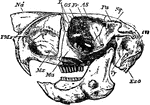
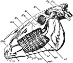
Cod Skull
"The skull of a cod. b, branchiostegal rays born on c.h., the ceratohyal bone; d, dentary portion of…
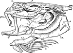
Cypris
"Cypris, side view, after removal of one valve. e., Eye; A.1, first antennae; A.2, second antennae;…
Dog Skull
"Longitudinal and Vertical section of the skull of a dog, with mandible and hyoid arch. an, anterior…
Horse Skull
"Side view of skull of horse, with the bone removed so as to expose the whole of the teeth. PMx, premaxilla;…
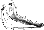
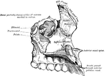
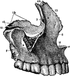
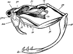
Inferior Maxilla of a Horse
Inferior maxilla of a horse-anterolateral view. Labels: a, body; b, b', rami; c, neck; d, mental foramen;…
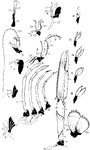
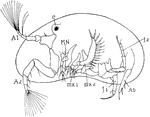
Norway Lobster Appendages
"Appendages of Norway lobster. Ex., Exopodite: En., endopodite; protopodite dark throughout; Ep., epipodite.…
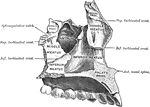
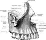
Maxilla
The largest bones of the face, excepting the mandible, and form, by their union, the whole of the upper…
Maxilla
The largest bones of the face, excepting the mandible, and form, by their union, the whole of the upper…

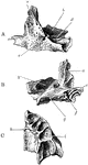
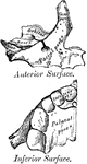
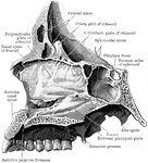
Ossification of Superior Maxilla
Ossification of superior maxilla. A, outer side. B, inner side. C, under side. Labels: a, nasal process;…
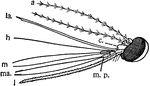
Mosquito Head
"The head of female mosquito (culex). a, antenna; c, clypeus; h, hypopharynx; m, mandibles; ma., maxillas;…
Proboscis
"Diagrammatic vertical section of the head and proboscis of a mosquito. l, labium bent as when the other…
Rattlesnake Skull
This illustration shows the skull of a rattlesnake. ar, articular portion of lower jaw; de, dentary…
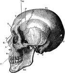
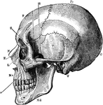
The Skull
A side view of the skull. Labels: O, occipital bone; T, temporal bone; Pr, parietal bone; F, frontal…
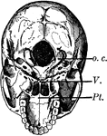
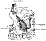
Base of the Skull
The base of the skull. The lower jaw has been removed. At the lower part of the figure is the hard palate…
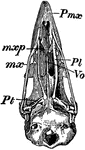
Bird Skull
"Schizognathous skull of common fowl. pmx, premaxilla; mxp, maxillopalatine; mx, maxilla; pl, palatine;…
Side View of the Skull
A side view of the skull. Labels: O, occipital bone; T, temporal; Pr, parietal; F, frontal; S, sphenoid;…
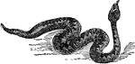
Common Viper
"Viper is a genus of venomous snakes. This family includes many important forms– e. g., the common…
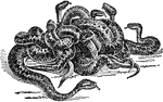
Common Vipers
"Viper is a genus of venomous snakes. This family includes many important forms– e. g., the common…