Clipart tagged: ‘pharynx’
Passage to the Esophagus and Windpipe
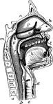
The passage to the esophagus and windpipe. Labels: c, the tongue; d, the soft palate ending in g, the…
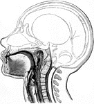
Head and Neck, Section of
Vertical middle section of head and neck showing the opening through the Eustachian tube, and its relations…
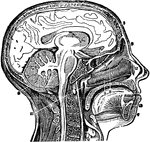
A Vertical Section of the Head and Neck
A vertical section of the head and neck through the mesial line, in order to show the opening of the…
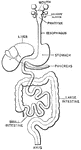
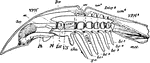
Digestive Apparatus of an Insect
"a, head, antennae, &c; b, pharynx; c, crop; d, gizzard; e, chyle-forming stomach; f, biliary vessels;…
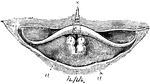
Larynx
The larynx viewed from its pharyngeal opening. The back wall of the pharynx has been divided and its…
Larynx
The larynx viewed from its pharyngeal opening. The back wall of the pharynx has been divided and its…
Upper Part of the Larynx
View of the upper part of the larynx as seen by means of the laryngoscope during the utterance of a…
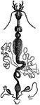
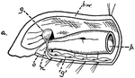
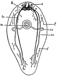
Limulus
"Diagram of a lateral view of a longitudinal section of Limulus. Suc, Suctorial pharynx. al, Alimentary…
Mouth
"The mouth, nose and pharynx, with the commencement of the gullet and larynx, as exposed by a section,…

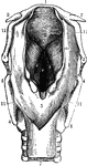
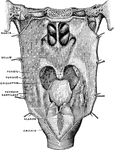
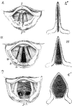
Horizontal Section Through Mouth and Pharynx
Horizontal section through mouth and pharynx at the level of the tonsils.
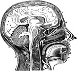
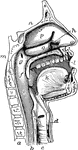
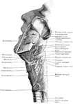
The Mouth, Nose, and Pharynx
The mouth, nose, and pharynx, with the commencement of gullet (esophagus) and larynx, as exposed by…
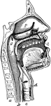
The Mouth, Nose, and Pharynx
The mouth, nose, and pharynx, with the larynx and commencement of gullet (esophagus), seen in section.…
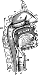
The Mouth, Nose, and Pharynx
The mouth, nose, and pharynx, with the commencement of the gullet (esophagus) and larynx, as exposed…
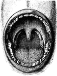
The Mouth
Interior of the mouth. Labels: 1, soft palate; 2, its median ridge; 3, uvula; 4, anterior, 5, posterior…
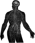
Nervous System
View of the nervous system in man, showing the nervous centers (the brain and the spinal near row) where…
Oligochete Worm
This illustration shows the arrangement of the nervous material in the anterior end of an Oligochete…
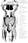
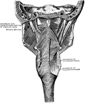
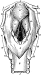
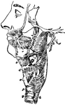
Muscles of the Pharynx
The muscles of the pharynx. On the right side most of the inferior constrictor has been removed, on…
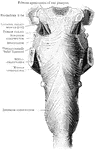
The Pharynx
The structure of the pharynx, a conical, musculo-membranous tube that forms the alimentary canal which…
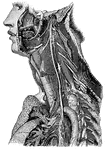
Trunk of the Pneumogastric Nerve
"Showing its distribution by its branches and ganglia to the larynx, pharynx, heart, lungs, and other…
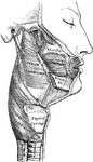
Swallowing Muscles
"The circular muscle of the mouth (1) and the buccinator or trumpeter's muscle (2) help the tongue to…
Turbellarian
This is a diagram of a Turbellarian, showing the general arrangement of the nervous structures and one…
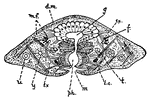
Turbellarian
This is a diagram of transverse section of a Turbellarian through the region of the mouth. d.m., dermo-muscular…
Movement of the Vocal Cords
Three laryngoscopic view of the superior aperture of the larynx and surrounding parts. Labels: A, the…