Clipart tagged: ‘ulna’
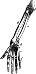
Bones of the Arm and Hand
Bones of the arm and hand. Labels: a, large end of ulna; b, radius; c, small end of the ulna; d, carpal…
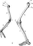
Arm and Leg Skeleton
The skeleton of the arm and leg. Labels: H, the humerus; Cd, its articular head which fits into the…
Arm Bones
"The bones of the arm. a, humerus; b, ulna; c, radius; d, the carpus; e, the fifth metacarpal; f, the…
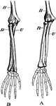
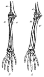
Bones of the Arm
Bones of the arm. Labels: A, arm in supination; B, arm in pronation. H, humerus; R, radius; U, ulna.
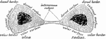
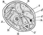
Transverse Section Through Arm
Transverse section through the middle of the right forearm, in the position of semipronation.
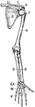
Bones of the Shoulder and Upper Extremity - Front View
"A, acromion; C, coracoid; CA, carpus; CL, clavicle; H, humerus; M, metacarpals; O, ventral surface…
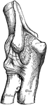
Elbow Joint
Right elbow joint, cut through at right angles to the axis of trochlea humeri, from the ulnar side.
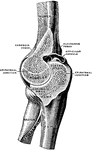
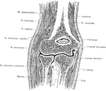
Frontal Section Through Elbow Joint
A view from behind of a frontal section through the right elbow joint.
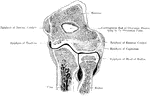
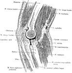
Sagittal Section Through Elbow Joint
A sagittal section through the left elbow joint of a child. View from the inner side.
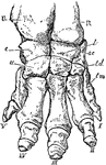
Elephant Foot
"Right fore foot of Indian Elephant. U, ulna; R, radius; c, cunelform; l, lunar; sc, scaphold; u, unciform;…
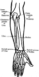
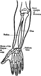
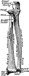
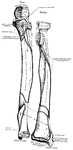
Forearm Bones
This diagram shows the bones of the right fore-arm. H, the humerus; R, the radius; and U, the ulna.
Forearm Bones
Transverse section through the bones of the forearm (radius and ulna), taken at about the middle of…
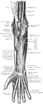
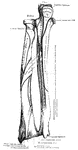
Muscles and Nerves of the Forearm
The muscles and nerves on the front of the forearm and hand. The pronator radii teres, flexor carpi…
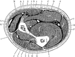
Section Across Forearm
Section across the forearm in the middle third. Labels: A, pronator radii teres; B, flexor carpi radialis;…
Forearm, Section of
A section across the forearm a short distance below the elbow-joint. R and U, its two supporting bones,…
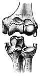
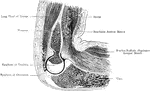
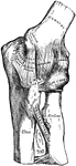
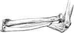
Elbow Joint
"Showing how the Ends of the Bones are shaped to form the Elbow Joint. The cut ends of a few ligaments…
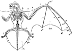
Noctule Bat
"Skeleton and volar Membranes of the Noctule Bat. c, clavicle; h, humerus; r, radius; u, ulna; d1, first…
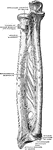
Ulna and Radius
"The ulna, or elbow bone, is the larger of these two bones. It is joined to the humerus by…
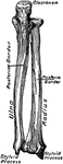
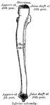
The Human Ulna and Radius
The Ulna and Radius. Labels: 1, radius; 2, ulna; o, olecranon process, on the anterior surface of which…
Fractured Ulna with Dislocation of Radius
Fracture of upper third of ulna, with dislocation of radius forward.
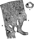
Transverse Section of Ulna
A, transverse section of the ulna, (bone of the arm), natural size, showing the medullary cavity. B,…
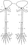
Vertebrate Appendages
"Diagrams of the girdles and appendages in a typical Vertebrate. A, anterior; B, posterior. ac., acetabulum,…
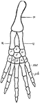
Vertebrate Fore Limb
"Ideal fore limb. H., Humerus; R., radius; U., ulna; r'., radiale; u'., ulnare; i., intermedium; c.,…
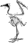
Vulture Skeleton
"Skeleton of Egyptian Vulture. (Neophron percnopterus), to show bones of bird. a, post-orbital process;…
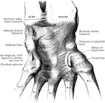
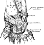
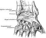
Ligaments of the Wrist
Ligaments on anterior aspect of radio carpal, carpal, and carpo metacarpal joints.