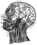
Muscles of the face, jaw and neck. 1, longus colli; 2, rapezius; 3, sterno-hyoid; 4, sterno-mastoid; 5, erico-thyroid; 6, trapezius; 7, constrictor of pharynx; 8, sterno-mastoid; 9, digastric; 10, attrahens aurem; 11, mylo-hyoid; 12, masseter; 13, depressor oof lower lip; 15, orbicularis oris; 17, levator of upper lip; 19, levator of angle of mouth; 21, orbicularis palpebrarum.

Facial Muscles

Muscles of the face, jaw and neck. 1, longus colli; 2, rapezius; 3, sterno-hyoid; 4, sterno-mastoid;…
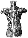
Muscles of the back. 1, trapezius; 2, its origin; 3, spine of scapula; 4, latissimus dorsi; 5, delltoid; 6, infra-spinatus; 7, external oblique; 8, gluteus medius; 9, gluteus maximus; 10, levator scapulae; 11, rhomboideus minor; 12, rhomboideus major; 13, splenius capitus; 14, splenius colli; 15, origin latissimus dorsi; 16, serratus inferior posticus; 17, supra-rpinatus; 18, infra-pinatus; 19, teres minor; 20, teres major; 21, long head triceps; 22, serratus major anticus; 23, internal oblique.

Back Muscles

Muscles of the back. 1, trapezius; 2, its origin; 3, spine of scapula; 4, latissimus dorsi; 5, delltoid;…
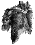
A frontal view of the chest muscles. 1, sterno-hyoid; 2, sterno-mastoid; 3, sterno-thyroid; 4, sterno-mastoid; 5, trapezius; 6, clavicle; 7, origin pectoralis major; 8, deltoid; 9, lower edge pectoralis major; 10, middle pectoralis major; 11, fibres external oblique; 12, biceps; 13, teres major; 14, serratus major anticus; 15, external oblique interlocking with serratus major.

Chest Muscles

A frontal view of the chest muscles. 1, sterno-hyoid; 2, sterno-mastoid; 3, sterno-thyroid; 4, sterno-mastoid;…
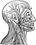
"(1) The facial nerve at its emergence from stylo-mastoid foramen; (2) temporal branches communicating with (3) the frontal branches of the fifth or trifacial nerve; (4) infraorbital branches communicating with (5) the infra-orbital branches of the fifth nerve; (6) maxillary branches communicating with (7) the mental branch of the fifth nerve; (8) cervico-facial branches; (9) the spinal accessory nerve giving off a branch to the trapezius muscles." — Winston's Encyclopedia, 1919

Facial Nerve

"(1) The facial nerve at its emergence from stylo-mastoid foramen; (2) temporal branches communicating…
Muscles of the arm

Arm Muscles

Muscles of the arm

Muscles of the upper body.

Muscles

Muscles of the upper body.

Muscles of the back.

Back Muscles

Muscles of the back.

Muscles of the arm.

Arm Muscles

Muscles of the arm.

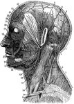
1. Temporal Artery 2. Artery behind the ear 3. Occipital Artery 4. Greater occipital nerve 5. Smaller ocipital nerve 6. Nerve of the neck 7. Trapezius muscle 8. Clavicular nerve 9. Clavicle 10. Sterno-0cleido-mastoid muscle 11.Outer artery of the head 12. Inner artery of the head 13. Salivary gland 14. Nerves of the lower jaw 15. Outer maxillary artery 16. Nerve of the chin 17. Circular muscle of th emouth 18. Greater yoke muscle 19. Nerves below the eye 20. Masseter or chewing muscle 21. Ear passage 22. Arteries of the forehead 23. Nerves of the forehead 24. Eye closing muscle 25.Facial Artery 26. Facial nerve

Neck

1. Temporal Artery 2. Artery behind the ear 3. Occipital Artery 4. Greater occipital nerve 5. Smaller…

Muscles of the back. Labels: 50, latissimus dorsi; 51, trapezius; 52, deltoid. The muscles of the back are in 5 layers, one beneath another. The two largest and most superficial are the trapezius and the latissimus dorsi.

Muscles of the Human Back

Muscles of the back. Labels: 50, latissimus dorsi; 51, trapezius; 52, deltoid. The muscles of the back…

Muscles of the back. Labels: 50, latissimus dorsi; 51, trapezius; 52, deltoid. The muscles of the back are in 5 layers, one beneath another. The two largest and most superficial are the trapezius and the latissimus dorsi.

Muscles of the Chest and Abdomen

Muscles of the back. Labels: 50, latissimus dorsi; 51, trapezius; 52, deltoid. The muscles of the back…
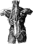
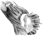
Second layer of muscles of the back. Labels: 1, trapezius; 2, a portion of the ten dinous ellipse formed by the trapezius on both sides; 3, spine of scapula; 4, latissimus dorsi; 5, deltoid; 6, infra-spinatus and teres minor; 7, external oblique; 8, gluteus medius; 9, gluteus magnus; 10, levator scapulae; 11, rhomboideus minor; 12, rhomboideus major; 13, splenius capitis; 14, splenius colli; 15, portion of origin of latissimus dorsi; 16, serratus inferior posticus; 17, supra-spinatus; 18, infra-spinatus; 19, teres minor; 20, teres major; 21, long head of triceps extensor cubiti; 22, serratus major anticus; 23, internal oblique.

Muscles of the Back

Second layer of muscles of the back. Labels: 1, trapezius; 2, a portion of the ten dinous ellipse formed…
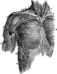
Superior muscles of the upper front of the trunk. Labels: 1, sterno-hyoid; 2, sterno-cleido-mastoid; 3, sterno-thyroid; 4, sterno-cleido-mastoid; 5, edge of the trapezius; 6, clavicle; 7, clavicular origin of the pectoralis major; 8, deltoid; 9, fold of the pectoralis major of the anterior edge of the axilla; 10, middle of the pectoralis major; 11, crossing and interlocking of the fibers of the external oblique of one side with those of the other; 12, biceps flexor cubiti; 13, teres major; 14, serratus major anticus; 15, superior heads of external oblique interlocking with serratus major.

Muscles of the Upper Trunk

Superior muscles of the upper front of the trunk. Labels: 1, sterno-hyoid; 2, sterno-cleido-mastoid;…
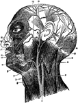
The arteries of the face and head. Labels: 1, common carotid; 2, internal carotid; 3, external carotid; 4, occipital artery; 5, superior thyroid artery; 6, trapezius; 7, lingual artery; 8, sterno-mastoid; 9, facial artery; 10, temporal artery, dividing into anterior and posterior branches; 11, submental branch; 12, transverse facial artery 13, inferior labial branch; 15, inferior coronary branch; 17, superior coronary branch; 19, lateral nasal branch; 21, angular branch.

Arteries of the Face and Head

The arteries of the face and head. Labels: 1, common carotid; 2, internal carotid; 3, external carotid;…
Arteries of the head. Labels: 1, common carotid; 2, internal carotid; 3, external carotid; 4, occipital; 5, superior thyroid; 6, trapezius; 7, lingual; 8, sterno-mastoid; 9, facial; 10, temporal; 11, submental; 12, transverse facial; 13, inferior labial; 15 inferior coronary; 17, superior coronary; 19, lateral nasal; 21, angular.

Arteries of the Head

Arteries of the head. Labels: 1, common carotid; 2, internal carotid; 3, external carotid; 4, occipital;…
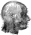
Nerves of the face and scalp. Labels: 1, attrahensaurem; 2, anterior belly occipito-frontalis; 3, auriculo-temporal nerve; 4, temporal branches of facial nerves; 5, attolens aurem; 6, supra-trochlea; 7, posterior belly occipito-frontalis; 8, supra-orbital; 9, retrahens aurem; 10, temporal branch of orbital nerve; 11, small occipital nerve; 12, malar branches of facial nerve; 13, posterior auricular nerve; 14, malar branch of orbital; 15, great occipital; 16, infra-orbital branches; 17, facial nerve; 18, nasal nerve; 19, cervico-facial division of facial; 20, infra-orbital branches; 17, facial nerve; 18, nasal nerve; 19, cervico-facial division of facial; 20, infra orbital nerve; 21, branches to digastric and stylo-hyoid; 22, temporo-facial division; 23, great auricular; 24, buccal branches of facial; 25, trapezius; 26, buccinator nerve; 27, splenius capitis; 28, masseter; 29, sterno-mastoid; 30, supra-maxillary branches of facial nerve; 31, superficial cervical nerve; 32, mental nerve; 33, platysma; 34, infra-maxillary branches of facial nerves.

Nerves of the Face and Scalp

Nerves of the face and scalp. Labels: 1, attrahensaurem; 2, anterior belly occipito-frontalis; 3, auriculo-temporal…
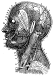
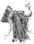
"Superficial arteries and nerves of the face and neck. 1, Temporal artery; 2, artery behind the ear; 3, occipital artery; 4, greater occipital nerve; 5, smaller occipital nerve; 6, nerve of the neck; 7, trapezius muscle; 8, clavicular nerves; 9, clavicle; 10, sterno-cleido-mastoid muscle; 11, outer artery of the head; 12, inner artery of the head; 13, salivary gland; 14, nerves of the lower jaw; 15, outer maxillary artery; 16, nerve of the chin; 17, circular muscle of the mouth; 18, greater yoke muscle; 19, nerves below the eye; 20, masseter, or chewing muscle; 21, ear passage; 22, arteries of the forehead; 23; nerves of the forehead; 24, eye-closing muscle; 25, facial artery; 26, facial nerve." -Foster, 1921

Arteries and Nerves

"Superficial arteries and nerves of the face and neck. 1, Temporal artery; 2, artery behind the ear;…

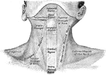
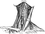
The region of the neck, from in front.

Front View of Neck

The region of the neck, from in front.

In mammals the muscles in their general plain resemble those of humans. The superficial muscles of a cow are diagramed. Labels: 1, Occipito-Frontalis. 2, Orbicularis Palpaebrarum. 3, Masseter. 5, Sterno-cleido-Mastoid. 6, Trapezius. 7, Latissimus Dorsi. 8, Pectoralis. 9, 10, External and Internal oblique muscles. 11, Opening of the mammary artery and vein (milk vein). 12, Gluteii. 13, Rectus Femoris muscle.

The Superficial Muscles of a Cow

In mammals the muscles in their general plain resemble those of humans. The superficial muscles of a…

In birds the muscles system is remarkable for their marked line of attachment to their tendons. Labels: 1, Occipito-Frontalis. 2, Orbicularis Palpaebrarum. 3, Temporal. 4, Masseter. 5, Sterno-cleido-Mastoid. 6, Trapezius. 7, Latissimus Dorsi. 8, Pectoralis. 9, Deltoid. 10, Biceps. 11, Triceps. 12, Gluteii. 13, Levator Caudae. 14, Rectus Femoris. 15, Gastrocnemius muscle.

The Superficial Muscles of a Hawk

In birds the muscles system is remarkable for their marked line of attachment to their tendons. Labels:…
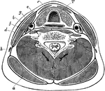
Transverse section through the lower part of the neck, to show the arrangement of the cervical fascia. Labels: a, Trapezius; b, sternomastoid; c, depressors of hyoid bone; d, platysma; e, anterior spinal muscles; f, scalenus anticus; g, carotid artery; h, external jugular vein; i, posterior spinal muscles; T, trachea, with gullet behind and thyroid body in front.

Transverse Section of the Neck

Transverse section through the lower part of the neck, to show the arrangement of the cervical fascia.…

Superficial and deep muscles of the trunk. The latissimus dorsi and trapezius on the right side have been cut away to expose the underlying muscles. Posterior View.

Posterior View of the Muscles of the Trunk

Superficial and deep muscles of the trunk. The latissimus dorsi and trapezius on the right side have…

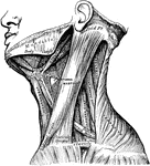
Muscles of the neck.

Neck Muscles

Muscles of the neck.

Anterior view of the muscles of the neck.

Anterior View of the Neck Muscles

Anterior view of the muscles of the neck.

Muscles of the back. On the left side is exposed the first layer; on the right side, the second layer and part of the third.

Back Muscles

Muscles of the back. On the left side is exposed the first layer; on the right side, the second layer…
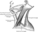
The triangles of the neck.

Triangles of the Neck

The triangles of the neck.

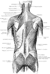
The superficial muscles of the back.

Superficial Muscles of the Back

The superficial muscles of the back.

Shown are the muscles of the deltoid region and back of the arm

Back View of Shoulder Muscles

Shown are the muscles of the deltoid region and back of the arm

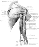
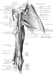
Shown are the muscles of the posterior wall of the axilla and the front of the arm (the biceps being divided).

Front View of Shoulder Muscles

Shown are the muscles of the posterior wall of the axilla and the front of the arm (the biceps being…

Scheme of the origin, connection, and distribution of the spinal accessory nerve. Labels: Sp.Acc, spinal accessory nerve; C.1-4, first four cervical nerves (dorsal roots); Va, pneumogastric nerve; R, ganglion of the root; t, ganglion of the trunk; G.Ph, glossopharyngeal nerve; S.M, nerves to sternocleidomastoid; Tr, nerves to trapezius; F.M, foramen magnum; J.F, jugular foramen.

Spinal Accessory Nerve

Scheme of the origin, connection, and distribution of the spinal accessory nerve. Labels: Sp.Acc, spinal…
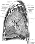
Sagittal section through left shoulder and left lung.

Sagittal Section Through Shoulder and Lung

Sagittal section through left shoulder and left lung.

Sagittal section through left shoulder, lung, and apex of the heart.

Sagittal Section Through Shoulder and Lung

Sagittal section through left shoulder, lung, and apex of the heart.
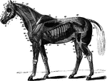
The superficial muscles of the horse with the panniculus and tunica abdominalis removed. Labels: 1, abducens; 2, retrahentes muscles; 2' attollens maximus; 2", attollens anticus; 3, temporalis; 4, nasalis longus; 5, orbicularis palpebrarum; 6, levator labii superioris alaeque nasi; 7, dilatator naris lateralis; 8, orbicularis oris; 9, zygomaticus; 9', buccinator; 10, depressor labii inferioris; 11, masseter; 12, levator humeri; 13, trapezius cervicalis; 14, trapezius dorsalis; 15, latissimus dorsi; 18, pextoralis parvus; 19, pectoralis magnus; 20, sterno-maxillaris; 23, subscapulo-hyoideus; 24, antea-spinatus; 25, teres externus; 26, postea-spinatus; 30, caput medium; 31, caput magnum; 33, extemsor metacarpi magnus; 34, humeralis obliquus; 35, extensor peis; 36, flexor metacarpi externus and medius; 37, flexor metacarpi internus; 46, cervical serratus magnus; 47, dorsal serratus magnus; 48, rhomboideus longus; 49, superficialis costarum; 50, splenius; 52, intercostales; 54, tensor fasciae latae; 55, triceps abductor femoris; 56, gluteus externus; 61, biceps rotator tibialis; 62, rectus femoris; 63, vastus externus; 65, gastrocnemius; 68, flexor pedis perforans; 69, peroneous; 70, extensor pedis; 71, flexor metatarsi; 72, flexor pedis accessorius; 74, obliquus abdominis externus.

Superficial Muscles of the Horse

The superficial muscles of the horse with the panniculus and tunica abdominalis removed. Labels: 1,…

"Muscles of a bird (accipiter nisus), after Carus, Tab. Anat. Comp., 1828, pl. 4.   a, pharynx; b, trachea; e, hyoid bone; d, ear; e, humerous; f, radius; g, ulna; h, radial finger; i, tibia; k, metatarsus; l, hind toe; m, inner toe; n, middle toe; o, outer toe. 1, biventer cervicis, with central tendon 1 a, and upper 1 b, and lower 1 c, belly. 2, complexus. 3, flexor capitis lateralis. 4, flexor longus capitis. 5, extensor magnus cervicis. 6, descendens cervicis. 7, 7, semispinales. 8, flexorsuperior capitis. 9, flexor inferior or longus capitis. 10, 10, intertransversales. 11, levator coccygis. 12, depressor coccygis. 13, cruro-coccygeus (ilio-coccygeus?). 14, pubo-coccygeus. 15 ischio-coccygeus. 16, lateralis quartus (quadratus coccygis, to tail-feathers). 17, obliquus externus abdominis. 18, cucullaris (trapezius). 19, serratus magnus. 20, pectoralis major. 21, a, b, latissimus dorsi. 22, deltoid. 23, suprascapular. 24, coraco-brachialis. 25, biceps brachii. 26, supinatpr longus. 27, anconeus longus (part of "triceps"). 28, anconeus brevis. 29, anconeus brevissimus. 30 a, 30 b, tensor patagii, carpal and radial parts. 31, tensor patagii posterior. 32, extensormetacarpi longus. 33, extensor metacarpi brevis. 34 a, flexor digitorum sublimis. 34 b, flexor digitorum profundus. 34 c, flexor metacarpi radialis. 36, flexor (meta-) carpi ulnaris. 37, glutaeus maximus. 38, adductor femoris primus. 39, sartorius. 40, latissimus femoris. 41, gracilis = ambiens: only its tendon in sight. 42, vastus; 43, iceps cruris. 44, semimembranosus. 46,46,47, gastrocnemius. 48 digastricus (chief opener of the mouth). 49, temporal. 50, long ligament. 51, cutaneous muscle of scalp. 52, masseter. 53, a muscle of the hyoid bone. 54, tibialis anticus. 55, tibialis posticus. 56, extensor hallucis. 57, flexor hallucis. 58, flaxor digitorum profundus or perforans, seen in various places: long and short head, and several tendons. 59, extensor longus digitorum, tendons seen in various places 60, abductor digiti interni. 61,61,61, flexores digitorum perforati. 62, peronaeus. 63, abductor minimi digiti. 64, abductor hallucis." Elliot Coues, 1884

Eurasian Sparrowhawk Muscles

"Muscles of a bird (accipiter nisus), after Carus, Tab. Anat. Comp., 1828, pl. 4. a, pharynx; b, trachea;…