Duck Skull

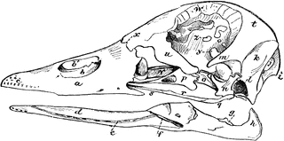
“Fig 63 - Skull of a duck (Clangula islandica), nat. size; Dr. R.W. Shufeldt, U.S.A. a, premaxillary bone; b, partly ossified internasal septum; b’, pervious part of nostril; c, end of premaxillary, perforated form numerous branches of second division of the fifth cranial nerve; d, dentary bone of under mandible; e, groove of nerves, etc.; f, a vacuity between dentary and other pieces of the mandible; g, articular surface; h, recurved “angle of the jaw;” i, occipital protuberance; j, vacuity in supraoccipital bone; k, muscular impression on back of skull; l is over the black ear-cavity; m, post-frontal process; n, quadrate bone; o, pterygoid; p, palatine; q, quadrato-jugal; r, jugal; s, maxillary; t, fronto-parietal dome of the brain-cavity; u; u, the lacrymal bone, immense in a duck, nearly completing rim of the orbit by approaching m; v, vomer; w, supra-orbital depression for the nasal gland; x, cranio-facial hinge; y, optic foramen; z, etc. interorbital vacuities.” Elliot Coues, 1884
Keywords
birds, duck, fowl, ornithology, bird anatomy, skull, bird bones, Clangula islandica, North American birds, internal parts of birds, fowl's skullGalleries
Bird AnatomySource
Elliot Coues Key to North American Birds (Boston, MA: Estes and Lauriat, 1884)
Downloads
2400×1196, 387.9 KiB
1024×510, 71.1 KiB
640×318, 36.9 KiB
320×159, 13.2 KiB