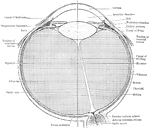
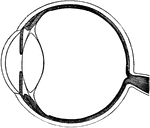
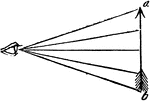
The Optics of the Eye ClipArt gallery includes 56 illustrations of the human eye. Additional views of the human eye are available in the Human Sensory Systems Clipart gallery under Human Anatomy.

"The angle under which the rays of light, coming from the extremities of an object, cross each other at the eye, bears a proportion directly to the length, and inversely to the distance of the object." -Comstock 1850

Angle of Vision

"The angle under which the rays of light, coming from the extremities of an object, cross each other…

An illustration depicting an astigmatism. An optical system with astigmatism is one where rays that propagate in two perpendicular planes have different foci. If an optical system with astigmatism is used to form an image of a cross, the vertical and horizontal lines will be in sharp focus at two different distances.

Diagram Illustrating Astigmatism

An illustration depicting an astigmatism. An optical system with astigmatism is one where rays that…

The convergence of light rays in the eyeball begins in the aqueous humor is perfected in the crystalline. But when they strike the vitreous humor, which is concave on its front surface, and also less dense than the crystalline, they diverse or spread out, and are thrown upon the back part of the eye.

Convergence of Rays in the Aqueous Humor of the Eyeball

The convergence of light rays in the eyeball begins in the aqueous humor is perfected in the crystalline.…
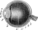
A section of the human eye.

Eye

A section of the human eye.

"Next in order is the aqueous humor, b, e, in the middle of which is the iris, d, c. Behind the pupil we have the crystalline lens, f, and then the vitreous humor, h, filling all the interior of the ball of the eye. m indicates the retina, which is an expansion of the optic nerve, n. k is the choroid coat, a membrane interposed between the retina and the sclerotic coat; it terminates in form in a series of folds or filaments, g, called the ciliary ligament or process." —Wells, 1857

Eye

"Next in order is the aqueous humor, b, e, in the middle of which is the iris, d, c. Behind the pupil…
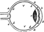
"a, sclerotic membrane; b, cornea; d, retina; o, optic nerve; v, vitreous humor." -Comstock 1850

Eye

"a, sclerotic membrane; b, cornea; d, retina; o, optic nerve; v, vitreous humor." -Comstock 1850
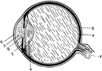
"The human eye-essential parts shown in section." —Croft 1917

Eye

"The human eye-essential parts shown in section." —Croft 1917

Section of the eye. 1: Optic nerve; 2: Retina; 3: Vitreous humor; 4: Crystalline lens; 5: Aqueous humor; 6: Cornea; 7: Iris; 8: Pupil.

Eye

Section of the eye. 1: Optic nerve; 2: Retina; 3: Vitreous humor; 4: Crystalline lens; 5: Aqueous humor;…
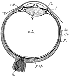
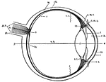
"Diagram of the eye. C., Cornea; a.h., aqueous humour; c.b., ciliary body; l., lens; I., iris; Sc., sclerotic; Ch., choroid; R., retina; v.h., vitreous humour; y.sp., yellow spot; n., optic nerve." -Thomson, 1916

Eye

"Diagram of the eye. C., Cornea; a.h., aqueous humour; c.b., ciliary body; l., lens; I., iris; Sc.,…
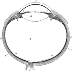
The human eye. Labels: a, crystalline lens; b, retina; c, cornea; d, sclerotic; e, choroid; g, ciliary processes; h, vitreous humor; k, pupil; i, iris; o, optic nerve.

Eye

The human eye. Labels: a, crystalline lens; b, retina; c, cornea; d, sclerotic; e, choroid; g, ciliary…
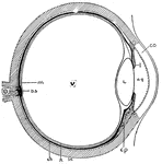
"Cross-section of the eye. Parts: co, cornea; I, iris; aq, anterior chamber of aqueous humor; L, lens; cp, ciliary process; sc, sclerotic coat; R, retina; ch, choroid; V, vitreous body; m, yellow spot; bs, blind spot; O, optic nerve." -Foster, 1921

Eye Cross-Section

"Cross-section of the eye. Parts: co, cornea; I, iris; aq, anterior chamber of aqueous humor; L, lens;…

Diagram of the eye. 1: Lines of light from end of arrow; 2: Small, inverted image in the eye.

Eye Diagram

Diagram of the eye. 1: Lines of light from end of arrow; 2: Small, inverted image in the eye.

The illustration of putting lenses in front of the eye. The focal point of the image is reflected into the eye. The focal point in the lens will end past the eye, creating a blurry image.

Optical Position and Size of Image Through Lens in Front of Eye

The illustration of putting lenses in front of the eye. The focal point of the image is reflected into…
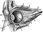
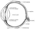
"Section through the left eye, closed. 1, lifting muscle; 2, upper straight muscle; 3, optic nerve; 4, fatty cushion; 5, lower straight muscle; 6, vitreous humor; 7, lower cross muscle; 8, lower eyelid; 9, upper eyelid; 10, crystalline lens." -Foster, 1921

Eye Section

"Section through the left eye, closed. 1, lifting muscle; 2, upper straight muscle; 3, optic nerve;…
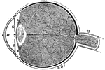
A section of the eye. Labels: 1, The sclerotic coat. 2, The cornea. 3, The choroid coat. 6, The iris. 7, The pupil. 8, The retina. 10, 11, Chambers or cavities of the eye that contain the aqueous humor. 12, The crystalline lens. 13, The vitreous humor. 15, The optic nerve. 14, 16, An artery of the eye.

A Section of the Eye

A section of the eye. Labels: 1, The sclerotic coat. 2, The cornea. 3, The choroid coat. 6, The iris.…

"...and the cornea will become too flat, or not suffciently convex, to make the rays of light meet at the proper place, and the image will therefore tend to be formed beyond the retina, instead of before it..." -Comstock 1850

Cornea too Concave on Eye

"...and the cornea will become too flat, or not suffciently convex, to make the rays of light meet at…

"If the cornea is too convex, or prominent, the image will be formed before it reaches the retina, for the same reason, that of two lenses, that which is most convex will have the least focal distance." -Comstock 1850

Cornea too Convex on Eye

"If the cornea is too convex, or prominent, the image will be formed before it reaches the retina, for…
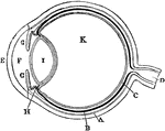
Plan of the eye seen in section. Labels: A, The Sclerotic Coat; B, The Choroid Coat; C, The Retina; D, The Optic Nerve; E, The Cornea; F, The Aqueous Humor; G, The Iris; H, The Ciliary Processes; I. The Crystalline Lens; K, The Vitreous Humor.

Diagram of the Eye

Plan of the eye seen in section. Labels: A, The Sclerotic Coat; B, The Choroid Coat; C, The Retina;…

"Diagram illustrating the Manner in which the Image of an Object is inverted on the Retina." — Blaisedell, 1904

Diagram of the Eye

"Diagram illustrating the Manner in which the Image of an Object is inverted on the Retina." — Blaisedell,…
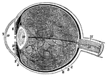
The right eye in horizontal section.

Horizontal Section of Eye

The right eye in horizontal section.

1, the sclerotic thicker behind than in front; 2, the cornea; 3, the choriod; 6, the iris; 7, the pupil; 8, the retina; 10, the anterior chamber of the eye; 11, the posterior chamber; 12, the crystalline lens, enclosed in its capsule; 13, the vitreous humour, enclosed in the hyaloid membrane and in cells formed in its interior by that membrae; 15, the sheath; and 16, the interior of the optic nerve, in the centre of which is a small artery.

Human Eye

1, the sclerotic thicker behind than in front; 2, the cornea; 3, the choriod; 6, the iris; 7, the pupil;…
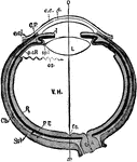
"Diagrammatic horizontal section of the eye of man. c, cornea; ch. choroid (dotted); C. P, ciliary processes; e. c, epithelium of cornea; e. cj, conjunctiva; f. c, yellow spot; I, iris; L, lens; ON, optic nerve; OS, ora serrata; o-x, optic axis; p. c. R, anterior non-visual portion of retina; P. E, pigmented epithelium (black); R, retina; sp. l, suspensory ligament; Scl, sclerotic; V. H, vitreous body." -Parker, 1900

Human Eye

"Diagrammatic horizontal section of the eye of man. c, cornea; ch. choroid (dotted); C. P, ciliary processes;…
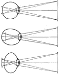
Diagrams of how an image is displayed with a normal eye (top image), myopic or nearsighted eye (middle image), and a hypermetropic or far-sighted eye. In a human, the retina "sees" an image upside down and the brain rights the image.

Human Eye

Diagrams of how an image is displayed with a normal eye (top image), myopic or nearsighted eye (middle…

"Section of the human eye." — Blaisedell, 1904

Human eye

"Section of the human eye." — Blaisedell, 1904

Lens of the eye. The rays of light are brought nearer together by the lenses of the eye, just as they are by the lenses of the telescope.

Lens of the Eye

Lens of the eye. The rays of light are brought nearer together by the lenses of the eye, just as they…

"Diagram showing the Change in the Lens during Accomadation. On the right the lens is arranged for distant vision, the cilliary muscle is relaxed, and the ligament <em>D</em> is tense, so flattening by its compression the front of the lens <em>C</em>, on the left the muscle <em>A</em> is acting, and this relaxes the ligament and allows the lens <em>B</em> to become more convex, and so fitted for the vision of near objects." — Blaisedell, 1904

Lens of the eye

"Diagram showing the Change in the Lens during Accomadation. On the right the lens is arranged for distant…
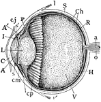
"Human Eye, in Median Vertical Anteroposterior Section. (Ciliary processes shown, through not all lying in this section.) A, anterior, and A', posterior chambers of aqueous humor; a, central artery of retina; C, cornea; Ch, choroid; cj, conjunctiva; cm, ciliary muscle; cp, ciliary processs; H, hyaloid; I, iris; L, crystalline lens in its capsule (the reference-line passes through the pupil); l, l', insertion of tendon of superior and inferior rectus muscles; o, optic nerve; P, canal of Petit; R, retina; S, sclerotic; s, s', circular sinus or canal of Schlemm; V, vitreous body filling back part of the eye." -Whitney, 1911

Median Vertical Anteroposterior Section of Eye

"Human Eye, in Median Vertical Anteroposterior Section. (Ciliary processes shown, through not all lying…

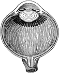
"Showing how the image of an object which is seen is formed on the retina of the eye." —Croft 1917
Eye Focusing on Object

"Showing how the image of an object which is seen is formed on the retina of the eye." —Croft 1917

Section of the eye magnified, showing the crystalline lens in its proper situation, between the aqueous and vitreous humors.

Section of the Eye

Section of the eye magnified, showing the crystalline lens in its proper situation, between the aqueous…
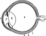
The eye. Labels: a, sclerotica; e, cornea; b, choroid; d, optic nerve; f, aqueous humor; g g , iris; h, lens; i, vitreous humor; c, retina.

The Eye

The eye. Labels: a, sclerotica; e, cornea; b, choroid; d, optic nerve; f, aqueous humor; g g , iris;…
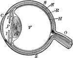
"The most essential parts of human vision are contained in the eyeball, a nearly spherical body, about an inch in diameter, and capable of being turned considerably in its socket by the action of various muscles." — Avery, 1895

Eyeball

"The most essential parts of human vision are contained in the eyeball, a nearly spherical body, about…
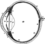
The left eyeball in horizontal section from before back. Labels: 1, sclerotic; 2, junction of sclerotic and cornea; 3, cornea; 4, 5, conjunctival membrane; 7, ciliary muscle; 10, choroid; 11, 13, ciliary processes; 14, iris; 15, retina; 16, optic nerve; 17, artery entering retina; 18, fovea centralis; 19, region where sensory part of retina ends; 26, 27, 28, are placed on the lens; 28 , suspensory ligament placed around lens; 29, vitreous humor; 30, aqueous humor in anterior chamber.

Left Eyeball in Horizontal Section

The left eyeball in horizontal section from before back. Labels: 1, sclerotic; 2, junction of sclerotic…
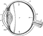
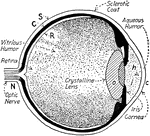
Section of the eyeball. Labels: Con, conjunctiva; C, cornea; A, aqueous humor; I, iris; L, crystalline lens; V, vitreous humor; Sc, sclerotic coat; Ch, choroid coat; R, retina; O, optic nerve, Cm, ciliary muscle.

Section of the Eyeball

Section of the eyeball. Labels: Con, conjunctiva; C, cornea; A, aqueous humor; I, iris; L, crystalline…
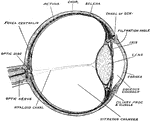
Horizontal section of the eyeball, showing the suspensory ligament of the lens, the aqueous and vitreous chambers, entrance of the optic nerve, and the fovea centralis.

Eyeball

Horizontal section of the eyeball, showing the suspensory ligament of the lens, the aqueous and vitreous…
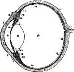
Diagram of a horizontal section through left eyeball and optic nerve.

Eyeball and Optic Nerve

Diagram of a horizontal section through left eyeball and optic nerve.

The left eyeball in horizontal section from before back. Labels: 1, sclerotic; 2, junction of sclerotic and cornea; 3, cornea; 4, 5, conjunctiva; 7, ciliary muscle; 10, choroid; 11, 13, ciliary processes; 14, iris; 15, retina; 16, optic nerve; 17, artery entering retina in optic nerve; 18, fovea centralis; 19, 20, region where sensory part of retina ends; 22, suspensory ligament; 24, the anterior part of the hyaloid membrane; 26, the lends; 29, vitreous humor; 30, aqueous humor.

The Eyeball in Horizontal Section

The left eyeball in horizontal section from before back. Labels: 1, sclerotic; 2, junction of sclerotic…
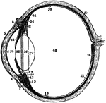
The left eyeball in horizontal section from before back. Labels: 1, sclerotic; 2, junction of sclerotic and cornea; 3, cornea; 4,5, conjunctiva; 6, posterior elastic layer of cornea; 7, ciliary muscle; 10, choroid; 11,13, ciliary processes; 14, iris; 15, retina; 16, optic nerve; 17, artery entering retina in optic nerve; 18, fovea centralis; 19, region where sensory part of retina ends; 22, suspensory ligament; 23 is placed in the canal of Petit, and the line from 25 points to it; 24, the anterior part of the hyaloid membrane; 26, 27, 28, are placed on the lends; 28 points to the line of attachment around it of the suspensory ligament; 29, vitreous humor; 30, anterior chamber of aqueous humor; 31, posterior chamber of aqueous humor.

Section of Left Eyeball

The left eyeball in horizontal section from before back. Labels: 1, sclerotic; 2, junction of sclerotic…

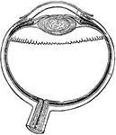
Vertical section of the eyeball.

Vertical Section of the Eyeball

Vertical section of the eyeball.

"A representation of the manner in which the image is formed in the eye, when the cornea or crystalline lens is flattened. The perfect image would be produced at m m, behind the retina, and, of course, beyond the point necessary to secure perfect vision." —Wells, 1857

Flattened Eye

"A representation of the manner in which the image is formed in the eye, when the cornea or crystalline…

Diagram to illustrate the mechanism of accommodation (focusing); on the right half of the figure for a near, on the left for a distant, object. Labels: rf, ciliary muscle; ch, ciliary process of choroid; sl, suspensory ligament; i, iris.

Focusing of the Eye

Diagram to illustrate the mechanism of accommodation (focusing); on the right half of the figure for…

In passing through the crystalline, the rays cross each other, so that those rays which pass from the lower part of an object are presented uppermost in the bottom of the eye and the reverse, so that the images of object are always inverted, or bottom side upwards. Labels: a, the aqueous; c, the crystalline; v, the vitreous humor.

Formation of an Image on the Eyeball

In passing through the crystalline, the rays cross each other, so that those rays which pass from the…

An illustration depicting the formation of circles of diffusion. "From point A luminous rays enter the eye in the form of a cone, the kind of which will depend on the pupil. Thus it may be circular, or oval, or ever triangular. If the pencil is focused in front of the retina, as at d, or behind it as it as at f, or, in other words, if the retina of being at F; be in the position G or H, there will be a luminous circle or a luminous triangular space, and many elements of the retina will be affected. The size of these diffusion circles depends on the distance from the retina of the point where the rays are focused: the greater the distance, the more extended will be the diffusion circle" (Britannica, 132).

Formation of Circles of Diffusion

An illustration depicting the formation of circles of diffusion. "From point A luminous rays enter the…
This diagram shows a side view of the right eye of man. a.c., central artery; a.h., aqueous humor; b., blind spot, the entrance of the optic nerve; c, conjunctiva; ch., choroid layer of the eye-ball; c.l., crystalline lens; c.m.c., circular fibers of the ciliary muscles; c.m.r., radial fibers of the ciliary muscles; co., cornea, the transparent portion of the sclerotic; c.p., ciliary process; c.s., canal of Schlemm, a lymphatic vessel; fo., fovea centralis, the point of clearest vision; o.n., optic nerve; o.s., ora serrata, the anterior wavy margin of the visual portion of the retina; r, the retinal layer; sc., sclerotic layer; sh., sheath of theoptic nerve; v.h., vitreous humor.

Human Eye

This diagram shows a side view of the right eye of man. a.c., central artery; a.h., aqueous humor; b.,…

"...where we suppose that the object a, is brought within an inch or two of the eye, and that the rays proceeding from it enter the pupil so obliquely as not to be refracted b the lens, so as to form a distinct image." -Comstock 1850

Indistinct Vision

"...where we suppose that the object a, is brought within an inch or two of the eye, and that the rays…

"The actual position of the vertical object a, as painted on the retina, is therefore such as is represented by the figure." -Comstock 1850

Inversion of Objects by the Eye

"The actual position of the vertical object a, as painted on the retina, is therefore such as is represented…

"Natural view of an arrow." -Comstock 1850

Natural Vision

"Natural view of an arrow." -Comstock 1850

"A representation of the manner in which the image is formed in the eye of a near-sighted person. The curvature of the cornea, s s, and of the crystalline lens, c c, is so great that the image is formed at m m in advance of the retina." —Wells, 1857

Near-sighted

"A representation of the manner in which the image is formed in the eye of a near-sighted person. The…

The lenses and humors of the eye must be very exactly arranged, in order that the sight may be perfect. They must be so arranged that the images of the objects shall be formed distinctly on the retina. In nearsighted persons the lenses and humors are so arranged as to make the rays that form the images from together too quickly, before they reach the retina, as shown in the figure.

Nearsighted Vision

The lenses and humors of the eye must be very exactly arranged, in order that the sight may be perfect.…

An illustration of the position and the size of the image viewed by the eye. The eye approximates the the height of the image. The image's height is the hypotenuse of the triangle, creating an angle with the eye. This image is then reflected upside down in the eye.

Position and Size of Image for Eye Optics

An illustration of the position and the size of the image viewed by the eye. The eye approximates the…

"The intersection of the principal rays in this case lies in the middle of the entrance pupil or of the exit pupil. By head and eye motion the various parts of the whole field can be viewed one after another." —Encyclopaedia Britannica, 1910

Optical Position of Diaphragms using Lens

"The intersection of the principal rays in this case lies in the middle of the entrance pupil or of…

"A representation of the manner in which the image is formed upon the retina in the perfect eye. The curvature of the cornea, s s, and of the crystalline lens, c c, is just sufficient to cause the rays of light proceeding from the image, e e, to converge the right focus, m, upon the retina." —Wells, 1857

Perfect Eye

"A representation of the manner in which the image is formed upon the retina in the perfect eye. The…

Vertical section of the pupil.

Vertical Section of the Pupil

Vertical section of the pupil.

"Diagram illustrating the points at which incident rays meet the retina. xx, optic axis; k, first nodal point; k', second nodal point; b, point where the image of B would be formed, were the eye properly accommodated for it; a, the retinal point where the image of A would be formed." —Martin, 1917

Retina

"Diagram illustrating the points at which incident rays meet the retina. xx, optic axis; k, first nodal…

A flattened shape of the globe of the eye causes farsightedness and an elongated shape of the globe of the eye causes nearsightedness. The diagram illustrates the position of the retina in these conditions. Labels: B, natural sight; G, farsightedness; C, nearsightedness.

The Position of the Retina in Near and Far Sight

A flattened shape of the globe of the eye causes farsightedness and an elongated shape of the globe…

Formation of an image in the retina.

Image Formation in the Retina

Formation of an image in the retina.

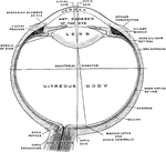
An illustration of a transverse section of an ideal eye. "A, summit of cornea; SC, sclerotic; S, Schlemm's canal; CH, choroid; I, iris; M, ciliary muscle; R, retina; N, optic nerve; HA, aqueous humour; L, crystalline lens, the anterior of the double lines on its face showing its form during accommodation; HV, vitreous humour; DN, internal rectus muscle YY', principle optical axis; C, [center] of the ocular globe..." (Britannica, 132).

Transverse Section of an Ideal Eye

An illustration of a transverse section of an ideal eye. "A, summit of cornea; SC, sclerotic; S, Schlemm's…