Human Connective Tissues
This human anatomy ClipArt gallery offers 54 illustrations of human connective tissues, including fibrous connective tissue (e.g., ligaments, tendons), cartilage, osseous tissue, and adipose tissue.

Achillles tendon
"Tendons are white, glistening cords, or straps, which connect the muscles with the bones." —…
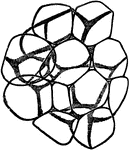
Adipose Tissue
Fat vesicles in adipose (fatty) tissue. Adipose tissue is a subtype of connective tissue consisting…
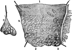
Adipose Tissue
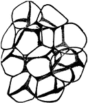
1. A portion of adipose (fat) tissue; 2. Minute bags containing the fat; 3. A cluster of the bags, separated…
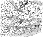
Adipose Tissue from Omentum
Adipose tissue from omentum. The fat cells are arranged as groups between the bundles of connective…

Cartilage Tissue
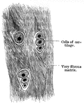
Fibrous cartilage connective tissue from the symphysis pubis, magnified. Cartilage is a structure without…

Longitudinal section of cartilage
"Showing (1) cartilage with martrix and cells; (2) cartilage with matrix containing cells and white…
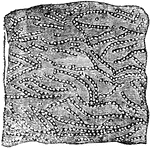
Cellular Tissue
A portion of cellular tissue, very highly magnified, showing the strings of globules of which its ultimate…

Connective Tissue
"Presently in the production of ordinay connective tissue, fibers of two kinds make their appearance…

Connective Tissue
"Presently in the production of ordinay connective tissue, fibers of two kinds make their appearance…

Connective Tissue
White fibrous tissue, a type of connective tissue composed of smaller fine wavy interlacing fibers which…

Connective Tissue
Yellow fibrous tissue, a type of connective tissue composed of larger fibers which branch and join each…
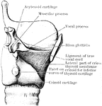
Cricothyroid Membrane
Dissection to show the lateral part of the cricothyroid membrane. The right ala of the thyroid cartilage…

Elbow-Joint
"The superficial veins in front of the elbow-joint. B', tendon of biceps muscle; Bi, brachialis internus…

Ciliated Epithelial Tissue
Ciliated epithelial tissue, which is covered with long waving hair-like projections (cilia). It is found…

Columnar Epithelial Tissue
Columnar epithelium lining a gland. It consists of conical cells laid side by side, their ends forming…
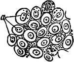
Pavement Epithelial Tissue
Pavement (flat) epithelium tissue, which is composed of flat scales, with nuclei of varying size and…

White fibrous tissue
"The connective tissue with white fibers sometimes forms a very thin sheet, as in the delicate covering…
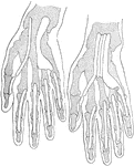
Tendon Sheaths of Wrist and Hand
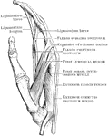
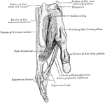
Projections of two types of flexor tendon sheaths. Note that in the hand upon the right side there is…
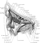
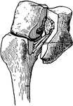
Inguinal Region and Hip Joint
Dissection of the structures beneath Poupart's ligament. The (*) indicates the iliopectineal intermuscular…
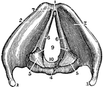
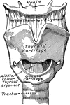
A View of the Larynx Showing the Vocal Ligaments
A view of the larynx showing the vocal ligaments. Labels: 1, The anterior edge of the larynx. 4, The…

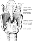
A Back View of the Cartilages and Ligaments of the Larynx
A back view of the cartilages and ligaments of the larynx. Labels: 1, The posterior face of the epiglottis.…

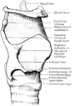
A Side View of the Cartilages of the Larynx
A side view of the cartilages of the larynx. Labels: *, The front side of the thyroid cartilage. 1,…

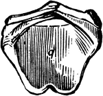
Arytenoid Cartilages of the Larynx
Arytenoid cartilages of the larynx. These are pitcher-like cartilages that are 2 in number, pyramidal-shaped,…

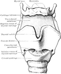
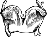
Cartilages from the Larynx
Cartilages from the larynx seen from the front. Labels: 1, vertical ridge of pomum Adami; 2, right ala;…
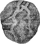
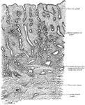
Mucous Membrane
A portion of the stomach, showing its internal surface or mucous coat. Mucous membranes line various…
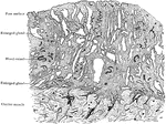
Mucous Membrane of Uterus in Fourth Month of Pregnancy
Section of mucous membrane lining body of uterus (decidua vera); fourth month of pregnancy.
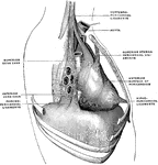
Pericardium Ligaments
The pericardium is a conical serofibrous sac in which the hear and the commencement of the great vessels…
Serous and Mucous Membranes
A diagram exhibiting the relative position of the elements of Serous and Mucous membranes. Labels: 1,…

Serous Membrane
A serious membrane. These line all closed cavities, or sac, of the body, and are reflected over the…
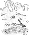
Synovial Membrane
Section of synovial membrane. Labels: a, endothelial covering of the elevations of the membrane; b,…
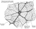
Transverse Section of a Tendon
Transverse section of a tendon, showing grouping of primary, secondary, and tertiary bundles of tendon…
Tendons of a Finger
The arrangement of the tendons of a finger. At a b c are the 3 bones of the finger. At f…
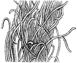
Yellow elastic tissue
"Along with white fibrous tissue, the yellow fibrous tissue... makes the coats of the arteries, and…
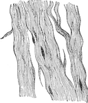
White Fibrous Tissue of the Tendon
Mature white fibrous tissue of tendon, consisting mainly of fibers with a few scattered fusiform cells.

