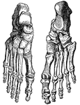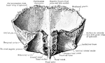
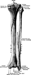
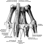
Bones of the Knee
Bones of the knee also showing muscle. Labels: s, insertion of the sartorius; g, insertion of the gracilis.
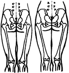
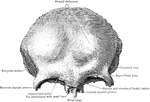
Position of the Pelvic and Thigh Bones in the Male and Female
Diagram showing the position of the pelvic and thigh bones in back view in the male and female.
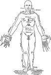
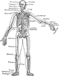
Front View of the Parts of the Human Body Labeled in English and Latin
Front view of a man in the anatomical position. On one lateral half the parts are labeled in English,…
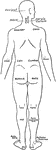
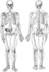
Back View of the Parts of the Human Body Labeled in English and Latin
Back view of a man. On one lateral the names of the parts are given in English, on the other in Latin.
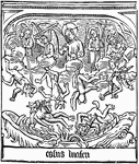
Speculum Humanae Salvationis
The engraved illustration of the Fall of Lucifer from the block book, Speculum Humanae Salvationis (Mirror…
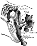
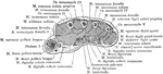
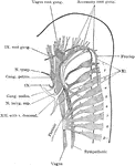
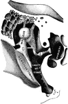
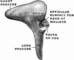
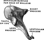
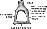
Small Bones and Ligaments of the Ear
Chains of small bones and their ligaments, seen from the front in a vertical, transverse section of…
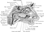
Nasal Cavity
The lateral wall of the left nasal cavity. The greater part of the middle turbinated bone has been excised…
Cross Section Through the Wrist Joint and Carpal Bones
Section through the wrist joint and carpal bones.
Cross Section Through Middle Metacarpal Bones of the Hand
Section through the middle of the right metacarpal bones.
Cross Section Through Distal End of the Metacarpal Bones of the Hand
Section through the distal end of the right metacarpal bones.
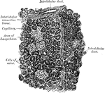
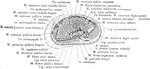
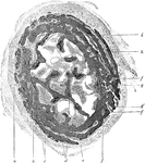
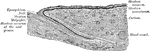
Section of Placenta
Section of human placenta at end of pregnancy. The fetal blood vessels have been injected; the maternal…
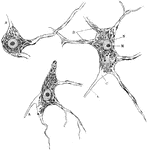
Neuron of Spinal Cord
Nerve cells of human spinal cord stained to show Nissl bodies. Labels: D, dendrites; A, axons; C, implantation…
Peripheral Nerves of Human Embryo
Reconstruction of peripheral nerves of human embryo of five weeks.
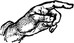
Hand Pointing
The hand is the intricate, multi-fingered body part normally located at the end of each arm of a human.
Transverse Section of Ureter
In human anatomy, the ureters are muscular tubes that propel urine from the kidneys to the urinary bladder.
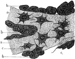
Developing Bone
Cross section of developing bone of human fetus of four months. Labels: a, periostem; b, boundary between…
Osteoblasts from Embryo
Osteoblasts from the parietal bone of a human embryo thirteen weeks old. Labels: a, bony septa with…
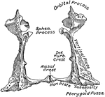
Internal Surface of Turbinated Bone
The turbinated bones are situated one on each side of the outer wall of each nasal fossa. Shown is the…
External Surface of Turbinated Bone
The turbinated bones are situated one on each side of the outer wall of each nasal fossa. Shown is the…
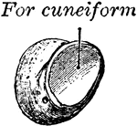
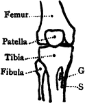
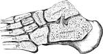
Pisiform Bone
The pisiform may be known by its small size and by its presenting a single articular facet. It is situated…
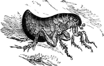
Human Flea
The human flea (Pulex irritans) is a parasitic insect that actually has several hosts despite its name.
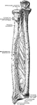
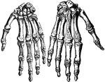
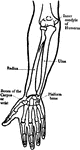
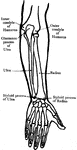
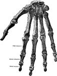
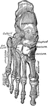
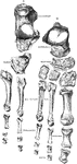
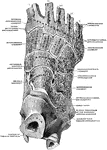
Bones of the Hand
Metacarpal bones and first phalanges of the second to the fifth of the right hand, with ligaments, from…
Metacarpal and Phalanges of Finger
Metacarpal bones and first phalanges of the third finger of the right hand, with ligaments, from the…
Microscopic view of a leaf
"The branch vascular bundles will be distinctly seen, resembling in some respects the arteries and veins…
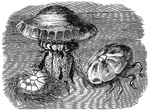
Rhyzostoma Cuvierii
"These fragile creatures are able to make long voyages on the surface of the sea. Their nature is such…
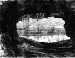
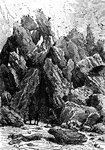
Cave
An illustration of a cliff with with a cave. A cave is a natural underground void large enough for a…
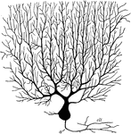
Purkinjean Cell from Cerebellum
Purkinjean cell from human cerebellum, as seen in a plane transverse to the long axis of a cerebellar…
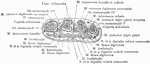
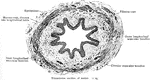
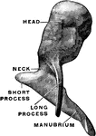
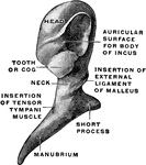
Ossicles of the Tympanum
Chain of ossicles and their ligaments, seen from the front in a vertical transverse section of the tympanum.
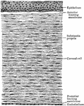
Longitudinal Section Through Fingernail
Longitudinal section through human nail and its nail groove (sulcus).
Transverse Section Through Fingernail
Transverse section through human nail and its nail groove (sulcus).