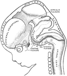
Profile view of brain of a human embryo of ten weeks. The various cranial nerves are indicated by numerals. A, Cerebral diverticulum of pituitary body. B, Buccal diverticulum of pituitary body.

Brain of Embryo

Profile view of brain of a human embryo of ten weeks. The various cranial nerves are indicated by numerals.…
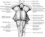
Front view of the medulla, pons, and mesencephalon of a full term human fetus.

Front view of Medulla, Pons, and Mesencephalon

Front view of the medulla, pons, and mesencephalon of a full term human fetus.
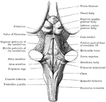
Back view of the medulla, pons, and mesencephalon of a full term human fetus.

Back View of Medulla, Pons, and Mesencephalon

Back view of the medulla, pons, and mesencephalon of a full term human fetus.
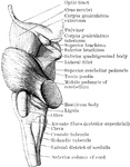
Lateral view of the medulla, pons, and mesencephalon of a full term human fetus.

Lateral View of Medulla, Pons, and Mesencephalon

Lateral view of the medulla, pons, and mesencephalon of a full term human fetus.
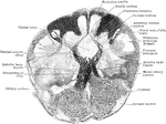
Transverse section through the closed part of the human medulla immediately above the decussation of the pyramids.

Transverse Section Through Closed Part of Medulla

Transverse section through the closed part of the human medulla immediately above the decussation of…
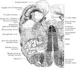
Transverse section through the human medulla in the lower olivary region.

Section Through Medulla in Olivary Region

Transverse section through the human medulla in the lower olivary region.

Transverse section through the the middle of the olivary region of the human medulla or bulb.

Section Through Medulla in Olivary Region

Transverse section through the the middle of the olivary region of the human medulla or bulb.
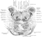
Section through the lower part of the human pons varolii immediately above the medulla.

Section Through Pons Varolii

Section through the lower part of the human pons varolii immediately above the medulla.
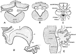
Showing the development of the cerebellum. A, Transverse section through the forepart of the cerebellum of a sheep embryo. B, Transverse section through the hinder part of the cerebellum of a sheep embryo. C, Cerebellum of a human fetus 17 cm long. D, Median section through cerebellum of sheep embryo 5 cm long.E, Median section through cerebellum of human fetus 17 cm long. Labels: 1, sulcus primarius; 2, sulcus above and afterwards behind the pyramid; 3, sulcus between pyramid and uvula; 4, great horizontal fissure; r.l., lateral recess ventricle IV; T, transverse groove in roof of ventricle IV.

Development of Cerebellum

Showing the development of the cerebellum. A, Transverse section through the forepart of the cerebellum…

An illustration of Biscayne Bay, is a lagoon that is approximately 35 miles (56 km) long and up to 8 miles (13 km) wide located on the Atlantic coast of south Florida. It is usually divided for purposes of discussion and analysis into three parts, North Bay, Central Bay and South Bay. North Bay separates Miami Beach on its barrier island from Miami on the mainland. It has been severely affected over the last century by raw sewage releases, urban runoff, shoreline bulkheading, dredging, the creation of artificial islands and the loss of natural fresh water flow into the bay. North Bay accounts for only 10% of the water area of the bay. Central Bay is the largest part of the bay. It is separated from the Atlantic Ocean by the Safety Valve, a series of shallow flats separated by tidal flow channels, stretching from the south end of Key Biscayne to the Ragged Keys at the north end of the Florida Keys. It has been adversely affected primarily by bulkheading, urban runoff discharged by canals, and the loss of natural fresh water flow. South Bay is nearly as large as Central Bay, and is the least affected by human activities, although it also suffers from the loss of natural fresh water flow. South Bay is separated from the Atlantic Ocean by the northernmost of the Florida Keys, and is connected to Florida Bay through channels and "sounds" lying between the mainland and the keys.

Biscayne Bay

An illustration of Biscayne Bay, is a lagoon that is approximately 35 miles (56 km) long and up to 8…
Daniel Coit Gilman (July 6, 1831-October 13, 1908) was an American educator.

Daniel Coit Gilman

Daniel Coit Gilman (July 6, 1831-October 13, 1908) was an American educator.

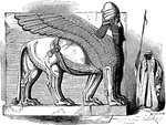
The Shedu is a celestial being from Mesopotamian mythology. He is a human above the waist and a bull below the waist. He also has the horns and the ears of a bull. The bull man helps people fight evil and chaos. He holds the gates of dawn open for the sun god Shamash and supports the sun disc. He is often shown on Cylinder Seals. It appears frequently in Mesopotamian art, sometimes with wings. Statues of the bull-man were often used as gatekeepers.

Shedu

The Shedu is a celestial being from Mesopotamian mythology. He is a human above the waist and a bull…
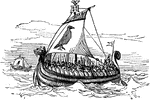
An illustration of a Norse Galley. Norse is an adjective relating things to Norway, Denmark, Faroe Islands, Greenland, Iceland and Sweden. A galley is an ancient ship which can be propelled entirely by human oarsmen, used for warfare and trade. Oars are known from at least the time of the Egyptian Old Kingdom. Many galleys had masts and sails for use when the winds were favorable.

Norse Galley

An illustration of a Norse Galley. Norse is an adjective relating things to Norway, Denmark, Faroe Islands,…
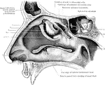
View of the outer wall of the nose. The turbinated bones having been removed. Labels: 1, vestibule, 2, hiatus semilunaris; 3, opening of antrum of highmore; 4, bulla ethmoidalis; 5, agger nasi; 6, opening of anterior ethmoidal cells; 7, cut edge of superior turbinated bone; 8, cut edge of middle turbinated bone; 9, pharyngeal orfice of Eustachian tube.

Outer Wall of Nose

View of the outer wall of the nose. The turbinated bones having been removed. Labels: 1, vestibule,…
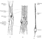
Olfactory and supporting cells in a frog and a human. A. Frog. B. Human. C. Human.

Olfactory and Supporting Cells

Olfactory and supporting cells in a frog and a human. A. Frog. B. Human. C. Human.

Diagrammatic section of the human retina.

Section of Retina

Diagrammatic section of the human retina.

A. A cone and two rods from the human retina (modified from Max Schultze); B. Outer part of rod separated into discs.

Cones and Rods of Retina

A. A cone and two rods from the human retina (modified from Max Schultze); B. Outer part of rod separated…
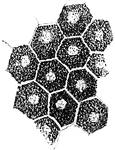
Pigment epithelium of human retina (viewed from the surface).

Pigment Epithelium of Retina

Pigment epithelium of human retina (viewed from the surface).

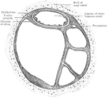
Transverse section of human semicircular canal.

Section of Semicircular Canals

Transverse section of human semicircular canal.

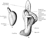
A, Left labyrinth of a human embryo of about four weeks; B, left labyrinth of a human embryo of about 5 weeks.

Development of Labyrinth

A, Left labyrinth of a human embryo of about four weeks; B, left labyrinth of a human embryo of about…

A, Section through papilla vallata of a human tongue. B, Section through part of the papilla foliata of a rabbit. Labels: 1, papilla; 2, vallum; 3, taste buds; 4, papillae; 5, taste buds; 6, ducts of serous gland.

Tongue of Human and Rabbit

A, Section through papilla vallata of a human tongue. B, Section through part of the papilla foliata…
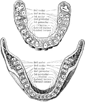
Horizontal section through both the upper and lower jaws to show the roots of the teeth. The sections were carried through the bones a short distance from the edge of their alveolar borders. The upper figure shows the upper teeth, the lower figure the lower teeth. Note the flattened roots of the lower incisors, the two root canals in the anterior root of each lower molar, and the confluence of the three roots of the upper wisdom teeth.

Jaw Showing Roots of Teeth

Horizontal section through both the upper and lower jaws to show the roots of the teeth. The sections…
An illustration of a woodpecker's skull.

Woodpecker Skull

An illustration of a woodpecker's skull.

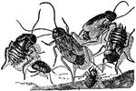
An illustration of a male (right) and female (left) cockroach. Cockroaches (or simply "roaches") are insects of the order Blattaria. This name derives from the Latin word for "cockroach", blatta. There are about 4,000 species of cockroach of which 30 species are associated with human habitations and about four species are well known as pests.

Cockroach

An illustration of a male (right) and female (left) cockroach. Cockroaches (or simply "roaches") are…

"Palmar aspect of left fore foot of a black bear (Ursus americanus). scl, scapholunar; c, cuneiform; p, pisiform; tr, trapezium; t, trapezoid; m, magnum; u, unciform. The phalanges show a full series of sesamoid bones (unmarked)." -Whitney, 1911

Bear Foot

"Palmar aspect of left fore foot of a black bear (Ursus americanus). scl, scapholunar; c, cuneiform;…
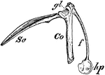
"Right shoulder-girdle or scapular arch of fowl, showing hp, the hypoclidium; f, furculum; Co, coracoid; Sc, scapula; gl, glenoid." -Whitney, 1911

Bird Scapula

"Right shoulder-girdle or scapular arch of fowl, showing hp, the hypoclidium; f, furculum; Co, coracoid;…

Watercresses are fast-growing, aquatic or semi-aquatic, perennial plants native from Europe to central Asia, and one of the oldest known leaf vegetables consumed by human beings. These plants are members of the Family Brassicaceae or cabbage family, botanically related to garden cress and mustard — all noteworthy for a peppery, tangy flavor.

Watercress

Watercresses are fast-growing, aquatic or semi-aquatic, perennial plants native from Europe to central…
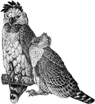
The American Harpy Eagle (Harpia harpyja) is a neotropical eagle, often simply called the Harpy Eagle. It is the only member of the genus Harpia. It is the largest and most powerful raptor found in the Americas, usually inhabiting tropical lowland rainforests in the upper (emergent) canopy layer. Its name references the harpies from Ancient Greek mythology. These were wind spirits that took the dead to Hades, and were said to have a body like an eagle and the face of a human.

American Harpy Eagle

The American Harpy Eagle (Harpia harpyja) is a neotropical eagle, often simply called the Harpy Eagle.…
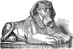
A Sphinx is a zoomorphic mythological figure which is depicted as a recumbent lion with a human head.
Sphinx

A Sphinx is a zoomorphic mythological figure which is depicted as a recumbent lion with a human head.

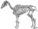
The skeleton of a horse. Axial Skeleton. The Skull. Cranial Bones: a, occipital, 1; b, wormian, 1; c, parietal, 2; d, frontal, 2; e, temporal, 2; sphenoid, 1; ethmoid, 1; auditory ossicles, 8. Facial bones: h, nasal, 2; g, lachrymal, 2; f, malar, 2; i, superior maxilla, 2; k, premaxila, 2; palatine, 2; pterygoid, 2; vomer, 1; tubinals, 4; l, inferior maxilla; 1; hyoid (segments), 5. Teeth: m, incisors, 12; n, canines, 4; o, molars, 24. The trunk: p-v, cervical vertebrae, 7; w, dorsal vertebrae, 18; x, lumbar vertebrae, 6; y, sacrum, 1; z, coccygeal vertebrae (variable), 18, d, d, ribs, 36; sternum (6-7 sternebrae), 7; *,coastal cartilages. Appendicular Skeleton. Pectoral Limb: e', scapula, 2; f', humerus, 2; g', radius, 2; h', ulna, 2. Carpus-i', trapesium, 2; k', cuneiform, 2; l', lunar, 2; m', scaphoid, 2; n', unciform, 2; o', magnum, 2; p', trapezoid, 2; q', pisiform, 2. Metacarpus-r', large bone, 2; s', small bone, 4. Digits- u', proximal phalanges, 2; v, median phalanges; w', distil phalanges, 2; t', large sesamoids, 4; small sesamoids, 2. Pelvic Limbs: Pelvis- b', ilium, 2; d' ischium, 2; c', pubis, 2. The limb- y', femur, 2; z', patella, 2; a', tibia, 2; b', fibula, 2. Tarsus- c', calcaneum, 2; d', astragalus, 2; e'. cuboid, 2; f', cuneiforme magnum, 2; g', cuneiforme medium, 2; h", cuneiforme parvum, 2. Metatarsus: large bone, 2; small bone, 4. Digit: proximal phalanges, 2; median phalanges, 2; distal phalanges, 2; large sesamoids, 4; small sesamoids, 2.

Skeleton of a Horse

The skeleton of a horse. Axial Skeleton. The Skull. Cranial Bones: a, occipital, 1; b, wormian, 1; c,…
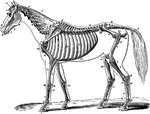
The skeleton of a horse. Showing its relation to the contour of the animal, viewed laterally. Labels: A, temporal fossa; B, inferior maxilla; C, atlas; D, dentata; E, cervical vertebrae; F, dorsal vertebrae; G, lumbar vertebrae; H, sacral vertebrae; I, coccygeal vertebrae; J, scapula; K, humerus; L, radius; L, ulna; M, carpus; N, trapezium; O, metacarpus; P, b. os suffraginis; Q, c. sesamoids; R, d. os coronae; S, e. os pedis; T, ribs; U., ilum; V, femur; X, patella; Y, tibia; y., Fibula; Z, tarsus; a, metatarsus; f, ligamentum nuchae, funicular portion; f' lamellar portion; 1, zygoma; 2, orbital fossa; 3, nasal peak; 4, incisor teeth; 4', canine teeth; 5, molar teeth; 6, external humeral trochanter; 7, scapular fossae; 8, coracoid apophysis; 9, cartilage of prolongation; 10, deltoid ridge, and external tuberosity; 11, olecranon; 12, costal cartilage; 13, anterior iliac spine; 14, ischium; 15, trochanter major; 16, trochanter minor; 18, anterior tibial tuberosity; 19, calcaneum; 20, small metacarpal and metatarsal or splint bones.

Skeleton of a Horse

The skeleton of a horse. Showing its relation to the contour of the animal, viewed laterally. Labels:…

"Schizognathous skull of common fowl. pmx, premaxilla; mxp, maxillopalatine; mx, maxilla; pl, palatine; pt, pterygoid; vo, vomer." -Whitney, 1911

Bird Skull

"Schizognathous skull of common fowl. pmx, premaxilla; mxp, maxillopalatine; mx, maxilla; pl, palatine;…

"Schizorhinal skull of curlew (top view), showing the long cleft, a, between upper and lower forks of each nasal bone." -Whitney, 1911

Curlew Skull

"Schizorhinal skull of curlew (top view), showing the long cleft, a, between upper and lower forks of…

Longitudinal section of a horse's skull. Labels: 1, supraoccipital bone and crest; 2, parietal bone; 3, frontal bone; 4, frontal sinus; 5, nasal bone; 6, superior turbinal; 12, inferior turbinal; 14, superior maxilla; 13 and its palatine plate; 15, premaxilla; 11, palatine bone; 7, ethmoid bone and volutes; 8, basi and presphenoid bones; 9, basioccipital bone; 10, petrosal bone. The cranium is the cavity under 1 and 2.

Longitudinal Section of Horse Skull

Longitudinal section of a horse's skull. Labels: 1, supraoccipital bone and crest; 2, parietal bone;…

The hyoid series is composed of five distinct pieces- a body, or hyoid bone proper, two cornua or horns and two cornicula or lesser horns. Shown us the left aspect of the hyoid series of bones in a horse. Labels: a, proximal, end of left cornu; b, distal end of right cornu; c, proximal end of corniculum; d, hyoid bone. The left heel process points downwards and backwards, the spur process in the opposite direction.

Hyoid Series of Bones in a Horse

The hyoid series is composed of five distinct pieces- a body, or hyoid bone proper, two cornua or horns…

External view of bones of right carpus, metacarpus, and digit of a horse. 1, distal end of radius; 2, groove for extensor tendons; 3, scaphoid; 4, lunar; 5, cuneiform;6, trapezium; 7, magnum; 8, unciform; 9, great metacarpal; 10, external small metacarpal (splint); 11, sesamoids; 12, proximal phalanx; 13, median phalanx; 5, distal phalanx; 16, basilar process.

Horse Leg

External view of bones of right carpus, metacarpus, and digit of a horse. 1, distal end of radius; 2,…

Posterior view of phalanges of a horse disarticulated. Labels: a, os suffraginis; b, os coronae; C, os pedis; D, os naviculare, or third sesamoid; 1, glenoid cavities joined by the groove, 2; 3, rough surface for V-shaped sesamoidean ligament; 4, condyles articulating with cavities, 5; 6, condyles of coronae articulating with glenoid cavities of pedal bone, 7; 8, pyramidal process; 9, basilar process; 10, retrosal processes; 11, articular surfaces between navicular and pedal bones; 12, surface, continues with glenoid cavities, 7; 13, posterior edge; 14, extremities for lateral ligaments of navicular bone.

Phalanges of a Horse

Posterior view of phalanges of a horse disarticulated. Labels: a, os suffraginis; b, os coronae; C,…

Bones of left tarsus of a horse, seen from in front and outside. Labels: 1, calcaneum; 2, astragalus; 3, cuneiforme magnum; 4, cuneiforme medium; 5, cuboid.

Tarsus of a Horse

Bones of left tarsus of a horse, seen from in front and outside. Labels: 1, calcaneum; 2, astragalus;…

External view of bones of left tarsus, metatarsus, and digit of a horse. Labels: 1, distal end of tibia; 2, calcaneum; 3, astragalus; 4, cuboid; 5, cuneiforme magnum; 6, cuneiforme medium; 7, great metatarsal; 8, external small metatarsal (splint); 9, proximal phalanx; 10, sesamoids; 11, median phalanx; 12, distal phlanx; 14, third sesamoid; 15, basilar process.

Horse Leg

External view of bones of left tarsus, metatarsus, and digit of a horse. Labels: 1, distal end of tibia;…
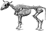
The skeleton of an ox. Axial Skeleton. The skull. Cranial Bones- occipital, 1: b, parietal, 2; a, frontal, 2; c, temporal, 2; sphenoid, 1; ethmoid, 1; auditory ossicles, 8. Facial Bones- h, nasal, 2; e, lachrymal, 2; d, malar, 2; f, maxilla, 2; g, premaxilla, 1; i, inferior maxilla, 2; palatine, 2; pterygoid, 2; vomer, 1; turbinals, 4; hyoid series, 7. Teeth: incisors, 6; canines, 2; molars, 24. The trunk- k, cervical vertebrae, 7; l, dorsal vertebrae, 13; m, lumbar vertebrae, 6; n, sacrum ( five segments), 1; o, coccygal vertebrae (variable), 20; p, ribs, 26; * sternum (seven sternebrae), 1. Appendicular skeleton. Pectoral limb. t, scapula, 2; u, humerus, 2; v, radius, 2; w, ulna, 2. Carpus- x, trapezium, 2; y, cuneiform, 2; z, lunar; a', scaphoid, 2; b', unciform, 2; c', magnum, 2. Metacarpus: d', large bone, 2; e', small bone, 4; f', large sesamoids, 8. Digit- g', proximal phalanges, 4; h', median phalages, 4; i', distal phalanges, 4; k', small sesamoids, 4. Pelvic Limb. Pelvis- q, ilium, 2; s, ischium, 2; r, pubis, 2. The limb- l', femur, 2; m', patella, 2; n', tibia, 2. Tarsus-o', maleolar, 2; p', calcaneum, 2; q', astragalus, 2; r', cubocuneiform, 2; s', cuneiforme medium, 2; t', cuneiforme parvum, 2. Metatarsus: large bone, 2; u', small bone, 2. Large sesamoids, 8. Digit: proximal phalanges, 4; median phalanges, 4; distal phalanges, 4; small sesamoids, 4. Visceral skeleton. Bones of the heart, 2. The separate bones of the ruminant skeleton, as here considered are 251.

Ox Skeleton

The skeleton of an ox. Axial Skeleton. The skull. Cranial Bones- occipital, 1: b, parietal, 2; a, frontal,…
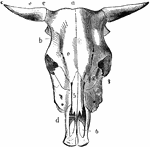
Skull of an ox, superior aspect. Labels: a, frontal crest; b, lateral crest; c, horn core; d, nasal peak; e, supraorbital foramina. Bones: 1, frontal; 2, lachrymal; 3, malar; 4, superior maxillary; 5, nasal; 6, premaxilla, with incisive openings.

Ox Skull

Skull of an ox, superior aspect. Labels: a, frontal crest; b, lateral crest; c, horn core; d, nasal…

Skeleton of the hog. Axial skeleton. The skull. Cranial bones- a, occipital, 1; b, parietal, 2; d, frontal, 2; c, temporal, 2; sphenoid, 1; ethmoid, 1; auditory ossicles, 8. Facial Bones- g, nasal, 2; h, os rostri, 1; f, lachrymal, 2; e, malar, 2; i, maxilla, 2; k, premaxilla, 2; l, inferior maxilla, 1; palatine, 2; 2, pterygoid, 2; vomer, 1; turbinals, 4; hyoid series, 5. Teeth: incisors, 12; canines, 4; molars, 28. The trunk- m, cervicle vertebre (variable), 18; r, ribs, 28; * sternum (seven sternebrae), 1. Appendicular Skeleton. Pectoral Limb. v, scapula, 2; w, humerus, 2; x, radius, 2; y, ulna, 2. carpus: z, trapezium, 2; a', cuneiform, 2; b', lunar, 2; c', scaphoid, 2; d', unciform, 2; e', magnum, 2; f', trapezoid, 2; g', pisiform, phalanges, 8; l', distal phalanges, 8; l", small sesamoids, 8. Pelvic Limb. Pelvis: s, ilium, 2; t, pubis, 2; u, ischium, 2. The limb-m', femur, 2; n', patella, 2; o', tibia, 2; p' fibula, 2. Tarsus- q' calcaneum, 3; r', astragalus, 2; s', cuboid, 2; t', cuneiforme magnum, 2; u', ecto-cuneiforme, 2; v' w', meso and endo cuneiformes, 4. Metatatarsus: large bones, 8; x', small bones 2; y', large sesamoids, 16. Digit: proximal phalanges, 8; median phalanges, 8; distal phalanges, 8; small sesamoids, 8. Visceral Skeleton- None. The separate bones of the skeleton of the hog, as here considered, are 234.

Skeleton of a Hog

Skeleton of the hog. Axial skeleton. The skull. Cranial bones- a, occipital, 1; b, parietal, 2; d, frontal,…
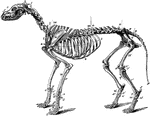
The skeleton of the dog. Axial skeleton. The skull. Cranial bones- a, occipital, 1; b, parietal, 2; d, frontal, 2; c, temporal, 2; sphenoid, 1; ethmoid, 2; auditory ossicles, 8. Facial Bones- f, nasal, 2; e, lachrymal, 2; d, malar, 2; h, maxilla, 2; g, premaxilla, 2. Teeth: incisors, 12; canines, 4; molars, 26. The trunk- l, cervical vertebrae, 7; m, dorsal vertebrea, 13; n, lumbar vertebrae, 7; o, sacrum (three segments), 1; p, coccygeal phalanges (variable), 20; t, ribs, 26; * sternum (eight sternebrae), 1. Appendicular Skeleton. Pectoral Limb. u, scapula, 2; v, humerus, 2; w, radius, 2; x, ulna. Carpus: y, trapezium, 2; z cuneiform, 2; a', scaphoid, 2; b', unciform, 2; c', magnum, 2; d', trapezoid, 2; e', pisiform,2; metacarpal bones, 10; h', anterior sesamoids, 10; g' posterior sesamoids, 20. Digits- i' proximal phalanges, 10; k', median phalanges, 8; l', distal phalanges, 10; small sesamoids wanting. Pelvic Limb. Pelvis: q, ilium, 2; r, pubis, 2; s, ischium, 2. The limb-m', femur, 2; o', fabellae, 4; n', patella, 2; q', tibia, 2; p', tibial sesamoid, 2; r', fibula, 2. Tarsus: s', calcaneum, 2; t', astragalus, 2; u', cuboid, 2; v', superior cuneiform, 2; w', ectocuneiforme, 2; x', mesocuneiforme, 2; y', edocuneiforme, 2. Metatarsus: large bones, 8; z',small bones, 2; anterior sesamoids, 8; posterior sesamoids, 16. Digit: proximal phalanges, 8; median phalanges, 8; distal phalanges, 8; small sesamoids wanting. Visceral Skeleton. Os Penis, 1; Rudimentary clavicle (inconstant), 2. The bones of the Carnivora Skeleton, thus considered, are 345.

Skeleton of a Dog

The skeleton of the dog. Axial skeleton. The skull. Cranial bones- a, occipital, 1; b, parietal, 2;…
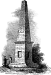
The Monument marks the grave site of the bones of victims of the Wyoming Massacre, which took place on July 3, 1778. Local residents banded together to defend the area against an invasion of British Tories as well as pro-Tory Native Americans. The battle ended in defeat for the colonial fighters and considerable brutality followed the actual Battle. In 1833, the bones were reinterred in a vault under the present Monument. The monument is located in the borough of Wyoming, Pennsylvania.

The Wyoming Monument

The Monument marks the grave site of the bones of victims of the Wyoming Massacre, which took place…
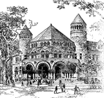
Many buildings stood on the Old Campus which were removed to make way for the current configuration of structures, including The Old Laboratory (1782–1888), The Cabinet (1819–1890), Trumbull Gallery (1832–1901), Alumni Hall (1853–1911, Alexander Jackson Davis; the towers from this building were rescued and rebuilt behind the tomb of Skull and Bones), Old Dwight Hall (1885–1926, J. Cleaveland Cady) and Osborn Hall (1888–1926, Bruce Price).

Osborn Hall, Yale University

Many buildings stood on the Old Campus which were removed to make way for the current configuration…

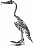
An illustration of a bird skeleton.

Bird Skeleton

An illustration of a bird skeleton.

An illustration of a fossil containing Chirotherium tracks. Chirotherium (also known as Cheirotherium) or 'hand-beast', is the name of a creature which may be known only from fossil imprints of its tracks (trace fossils). These look, by coincidence, remarkably like the hand of an ape/human or bear, with the outermost toe having evolved to extend out to the side like a thumb, although probably only providing a firmer grip in mud. Its tracks were first found in 1834, in red sandstone in Thuringia, Germany, dating from 240 million years ago (mya). This creature was probably an archosaur, related to the ancestors of the dinosaurs.

Chirotherium Tracks

An illustration of a fossil containing Chirotherium tracks. Chirotherium (also known as Cheirotherium)…
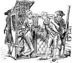
The sedan or litter is a wheelless, human-powered vehicle used to carry one person sitting inside.
Sedan

The sedan or litter is a wheelless, human-powered vehicle used to carry one person sitting inside.

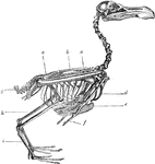
The skeleton of a bird. Labels: a, radius and ulna; b, dorsal vertebrae; c, sacrum and pelvis; g, ploughshare bone; h, tibia; i, metatarsus; d, furculum; e, carina of the sternum; f, distal phalanx of pectoral limb.

Skeleton of a Bird

The skeleton of a bird. Labels: a, radius and ulna; b, dorsal vertebrae; c, sacrum and pelvis; g, ploughshare…
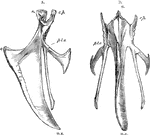
The sternum of a bird. Label: a, lateral aspect; b, inferior aspect; r, rostrum; c.p, costal process; pl.o, pleurosteon; c, carina; m.x, middle xiphoid process.

Sternum of a Bird

The sternum of a bird. Label: a, lateral aspect; b, inferior aspect; r, rostrum; c.p, costal process;…
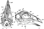
The skull of a bird. Labels: a, inferior aspect, the mandible being removed; b, lateral aspect; px, premaxilla; mx, superior maxilla; p, palatine bone; v, vomer; j, jugal bone; qj, quadrato-jugal bone; lachrymal; me, median ethmoid; f, frontal; pf, post-frontal process; p, parietal; pt, pterygoid; q, os quadratum; sq, sqamosal; eu, process before the sustachian aperture; bo, basi-occipital, and so, supra-occipital bone; ar, articular; a, surangular, and d, dentary portion of the mandible.

Skull of a Bird

The skull of a bird. Labels: a, inferior aspect, the mandible being removed; b, lateral aspect; px,…
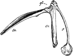
The pectoral arch of a bird. Labels: sc, scapula; co, coracoid bone; f, clavicle, terminating below in the hypocleideum; gl, glenoid cavity.

Pectoral Arch of a Bird

The pectoral arch of a bird. Labels: sc, scapula; co, coracoid bone; f, clavicle, terminating below…
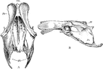
The pelvis of a bird. Labels: a, superior; b, lateral aspect; sm, sacrum; Il, ilium; Is, ischium; Am, acetabulum.

Pelvis of a Bird

The pelvis of a bird. Labels: a, superior; b, lateral aspect; sm, sacrum; Il, ilium; Is, ischium; Am,…
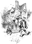
A pixy named Thomas alarmed that a human has just invaded his lawn by jumping over Thomas' wall.

Pixy and a Man

A pixy named Thomas alarmed that a human has just invaded his lawn by jumping over Thomas' wall.

Pogonias chromis, a name of several fish so called from the drumming noise they make, said to be due, in part at least, to the grinding of bones upon each other.

Saltwater Drum

Pogonias chromis, a name of several fish so called from the drumming noise they make, said to be due,…

An illustration of: 1, Coralla cut open showing the four stamens; 2, Unripe fruit (lengthwise); 3, ripe capsule split open; and the foxglove plant. Digitalis is a genus of about 20 species of herbaceous perennials, shrubs, and biennials that are commonly called foxgloves. The genus was traditionally placed in the figwort family Scrophulariaceae, but upon review of phylogenetic research, it has now been placed in the much enlarged family Plantaginaceae. The genus is native to Europe, western and central Asia, and northwestern Africa. The scientific name means "finger-like" and refers to the ease with which a flower of Digitalis purpurea can be fitted over a human fingertip. The flowers are produced on a tall spike, are tubular, and vary in colour with species, from purple to pink, white, and yellow. The best-known species is the Common Foxglove, Digitalis purpurea. It is a biennial, often grown as an ornamental plant due to its showy flowers, that range in colour from purples through to whites, with variable marks and spotting. The first year of growth produces only the long, basal leaves. In the second year, the erect leafy stem 0.5-2.5 m tall develops.

Foxglove

An illustration of: 1, Coralla cut open showing the four stamens; 2, Unripe fruit (lengthwise); 3, ripe…

Corystes cassivelaunus, the masked crab, helmet crab or sand crab, is a burrowing crab of the North Atlantic and North Sea from Portugal to Norway, which also occurs in the Mediterranean Sea. It may grow up to 4 cm long (carapace length). The name "masked crab" derives from the patterns on the carapace which resemble a human face, in a similar manner to heikegani.

Masked Crab

Corystes cassivelaunus, the masked crab, helmet crab or sand crab, is a burrowing crab of the North…
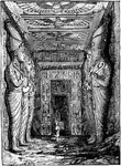
In 1959 an international donations campaign to save the monuments of Nubia began: the southernmost relics of this ancient human civilization were under threat from the rising waters of the Nile that were about to result from the construction of the Aswan High Dam. This image gives a perspective view of the entrance hall, in which are seen eight standing colossal statues 33 feet high, supported by square piers.1

Entrance to the Great Temple at Abu Simbel

In 1959 an international donations campaign to save the monuments of Nubia began: the southernmost relics…
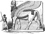
The Sumerian word lama, which is rendered in Akkadian as lamassu, refers to a beneficient protective female deity. The corresponding male deity was called alad, in Akkadian, šêdu. In art they were depicted as hybrids, as winged bulls or lions with the head of a human male (Centauroid). There are still surviving figures of šêdu in bas-relief and some statues in museums. Notable examples of šêdu/lamassu held by museums include those at the British Museum, Musée du Louvre, National Museum of Iraq, Metropolitan Museum of Art and one extremely large example kept at the Oriental Institute, Chicago. They are generally attributed to the ancient Assyrians.

Winged Bull from Nimrud

The Sumerian word lama, which is rendered in Akkadian as lamassu, refers to a beneficient protective…
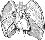
The human thoracic viscera.

Thoracic Viscera

The human thoracic viscera.