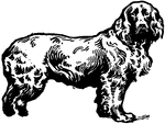
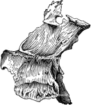
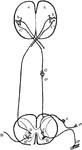
Field Spaniel
"The field spaniel, a dog to which very great attention has been paid by breeders and fanciers, who…
Eye-splice
"For making an eye splice, the end of the rope is unlaid and the strands are bent upon the body of the…
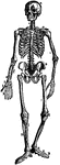
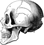
The Human Skeleton
The Human Skeleton. Labels: a, parietal bone; b, frontal; c, cervical vertebrae; d, sternum; e, lumbar…
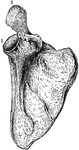
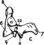
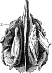
The Human Scapula
The human scapula bone (shoulder blade). Labels: 1, glenoid cavity; 2, end of the spine of scapula.
The Human Humerus
The human humerus bone, the longest and largest bone of the upper leg. Labels: a, rounded head; gt,…
The Human Ulna and Radius
The Ulna and Radius. Labels: 1, radius; 2, ulna; o, olecranon process, on the anterior surface of which…
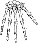
The Human Wrist and Hand Bones
Bones of the Wrist and Hand. Labels: m, metacarpal bones; p, phalanges; 3, bones of wrist.

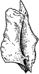
Part of the Human Pelvic Bone
The Os Innominatum, or nameless bone, so called from bearing no resemblance to any known object, is…
Human Femur Bone
The Femur (upper leg bone) is the longest, largest, and strongest bone in the skeleton. Labels: b, rounded…
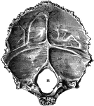
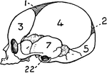
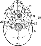
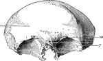
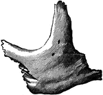
Occipital Bone of the Human Skull
Occipital bone of the human skull, inner surface. It is situated at the back and base of the skull.…
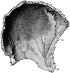
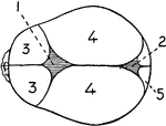
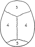
Parietal Bone of the Human Skull
Parietal bone of the human skull, inner surface. The parietal bones form the greater part of the sides…
Frontal Bone of the Human Skull
Frontal bone of the human skull, outer surface. The frontal bone forms the forehead, roof of the orbital…
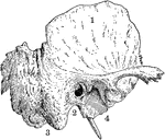
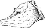
Temporal Bone of the Human Skull
Temporal bone of the human skull. The temporal bones are situated at the sides and base of the skull.…
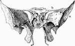
Sphenoid Bone of the Human Skull
Sphenoid bone, situated the anterior part of the base of the skull, articulating with all the other…
Ethmoid Bone of the Human Skull
Ethmoid bone, posterior surface. The ethmoid bone is an exceedingly light, spongy bone, placed between…
Human Lachrymal Facial Bone
Lachrymal Bone. The lachrymal are the smallest and most fragile bones fo the face. They are situated…
Human Vomer Nasal Bone
Vomer bone, a single bone placed at the back part of the nasal cavity, and forms part of the septum…
Human Malar (Cheek) Bone
Malar (cheek) bone. The malar bones form the prominence of the cheek, and part of the outer wall and…
Human Palate Bone
Palate bone. Palate bones form the back part of the roof of the mouth; part of the floor and outer wall…
Human Nostril Bone
Inferior turbinated bone, convex surface. The inferior turbinated bones are situated on the outer wall…
Human Maxillary (Upper Jaw) Bone
Superior maxillary bone. With it's fellow on the opposite side, it forms the whole of the upper jaw.…
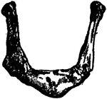
Human Maxillary (Upper Jaw) Bone
Inferior Maxillary Bone (lower jaw). It is the largest and strongest bone in the face and serves for…
Human Hyoid Bone
The hyoid, os hyoides, or tongue bone, is an isolated, U-shaped bone lying in front of the throat, just…
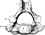
Human Cervical Vertebra Bone
A cervical vertebra of the spine, inferior surface. Labels: 1, spinous process, slightly bifid; 4, transverse…
Human Spinal Column
Side view of spinal column, without sacrum and coccyx. Labels: 1 to 7, cervical vertebrae; 8 to 19,…
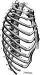
Human Thorax (Chest)
Thorax. The thorax, or chest, is an elongated conical-shaped cage, formed by the sternum and costal…
Human Sternum Bone
Sternum, front and side view. The sternum, or breast bone, is a flat narrow bone, situated in the median…
Human Skull
The skull. Labels: a, nasal bone; b, superior maxillary; c, inferior maxillary; d, occipital; e, temporal;…
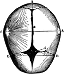
Human Skull at Birth
The skull at birth, superior suerface. The cranial bones of the infant at birth are not fullyformed…
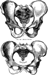
Human Pelvis, Male and Female
Male pelvis (top) and female pelvis (bottom). The pelvis is stronger and more massively constructed…
Human Joint, Mixed Articulation
A mixed articulation (slightly movable). In this form, the bony surfaces are usually joined together…
Human Joint, Dentated Suture
A toothed, or dentated suture. This is one type of immovable articulation. It is found in the union…
Muscle Fiber
Diagram of muscle fiber with sarcolemma attached. Muscular tissue is the tissue by means of which the…
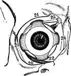
Muscles of the Human Eyeball
Muscles of right eyeball within the orbit, seen from the front. Labels: 21, superior rectus; 22, inferior…
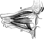
Muscles of the Human Eyeball
Muscles of eyeball, seen from side. Labels: 19, elevator muscle of eyelid; 22, inferior rectus; 23,…
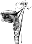
Muscles of the Human Tongue
Muscles of the tongue. The chief muscles connecting the tongue and tongue bone to the lower jaw are…
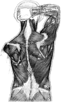
Muscles of the Human Back
Muscles of the back. Labels: 50, latissimus dorsi; 51, trapezius; 52, deltoid. The muscles of the back…
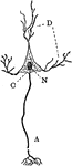
Diagram of a Neuron
Diagram of a neuron. Labels: A, axon arising from the cell-body and branching at its termination; D,…
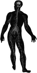
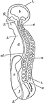
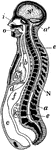
Diagram of the Human Nervous System
Diagram illustrating the general arrangement of the cerebrospinal nervous system.
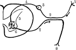
Nerve Reflex Arc
Reflex arc, as it is approximately in man. Labels: 1, nerve terminal, or sensory epithelium; 2, dendrite…
Nervous System Diagram
Diagram of nervous system. Labels: a, a, cortex of cerebral hemispheres; b, b, cell body and dendrites…
!["A verticle section of one form of blast-furnace is represented [here]. the crucible (C) is the part of the furnace in which the molten matte and slag collect. The body of the furnace consists of two concentric shells (cs), made either of wrought iron or of steel, between which cold water (W) is caused to circulate to precent the inner shell becoming heated...Pipes called tuyeres (T) enter the furnace a short distance above the hearth...Above the body of the furnace extend the hood (H) and the stack (S). A door (D), used in charging the furnace, is placed in the hood." -Brownlee 1907](https://etc.usf.edu/clipart/35400/35491/blast_fur_35491_mth.gif)
Blast-furnace
"A verticle section of one form of blast-furnace is represented [here]. the crucible (C) is the part…
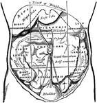
Regions of the Abdomen and their Contents
Regions of the abdomen and their contents (edge of costal cartilages in dotted outline). "For convenience…
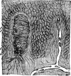
Section of Epidermis (Skin)
Section of skin showing two papillae and deeper layers of epidermis. The papillae contain blood vessels…
Piece of Human Hair
Piece of human hair, highly magnified. Labels: a, cuticle; b, fibrous substance; c, medulla.
Trunk and Head of Human Body
Diagrammatic longitudinal section of the trunk and head. Labels: 1,1, the dorsal cavity; a, the spinal…
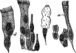
Ciliated Epithelium Cells
Ciliated epithelium from the human trachea, highly magnified. Labels: a, large ciliated cell; d, cell,…
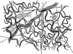
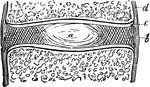
Subcutaneous Areolar Tissue from a Young Rabbit
Subcutaneous areolar tissue from a young rabbit, highly magnified. The white fibers are in wavy bundles,…

Fibrous Tissue of a Tendon
Fibrous tissue, from the longitudinal section of a tendon. "The fibrous tissue is met with in the form…
Longitudinal Section of Body
Diagrammatic longitudinal section of the body. Labels: a, the neural tube, with its upper enlargement…
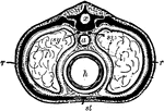
Section Across the Body in the Chest Region
A Diagrammatic section across the body in the chest region. Labels: x, the dorsal tube, which contains…
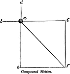
Compound Motion
"Compound motion is that which is produced by two or more forces, acting in different directions, on…