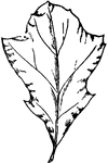
The leaf of a Black oak tree.

Black Oak Leaf

The leaf of a Black oak tree.

The leaf of a laurel-oak tree.

Laurel-Oak Leaf

The leaf of a laurel-oak tree.

The leaf of a willow-oak tree.

Willow-Oak Leaf

The leaf of a willow-oak tree.

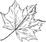
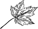
The leaf of a Norway Maple tree.

Norway Maple

The leaf of a Norway Maple tree.

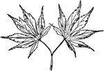
The leaf of a Japanese Maple tree.

Japanese Maple

The leaf of a Japanese Maple tree.

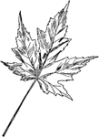
The leaf of a Silver Maple tree.

Silver Maple

The leaf of a Silver Maple tree.

The leaf of a Red or Swamp Maple tree.

Red or Swamp Maple

The leaf of a Red or Swamp Maple tree.

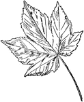
The leaf of a Sycamore Maple tree.

Sycamore Maple

The leaf of a Sycamore Maple tree.

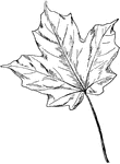
The leaf of a Sugar Maple tree.

Sugar Maple

The leaf of a Sugar Maple tree.

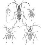
"The 'leaf bug' (Dicyphus minimus): a, newly hatched; b, second stage; c, nymph; d, adult; e, head and beak from side--enlarged (original)." -Department of Agriculture, 1899

Leaf Bug

"The 'leaf bug' (Dicyphus minimus): a, newly hatched; b, second stage; c, nymph; d, adult; e, head and…

A leaf damaged by the tobacco split worm.

Split Worm Damage

A leaf damaged by the tobacco split worm.

The larva and pupa stages of the cabbage plusia.

Cabbage Plusia Larva and Pupa

The larva and pupa stages of the cabbage plusia.

The larva of the bean leaf-beetle, Cerotoma trifurcata.

Bean Leaf-Beetle Larva

The larva of the bean leaf-beetle, Cerotoma trifurcata.

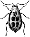
The adult of the bean leaf-beetle, Cerotoma trifurcata.

Bean Leaf-Beetle

The adult of the bean leaf-beetle, Cerotoma trifurcata.

The pupa of the bean leaf-beetle, Cerotoma trifurcata.

Bean Leaf-Beetle Pupa

The pupa of the bean leaf-beetle, Cerotoma trifurcata.

The leaves of the common rose geranium, Pelargonium capitatum.

Rose Germanium

The leaves of the common rose geranium, Pelargonium capitatum.

The leaves of the skeleton-leaved geranium, Pelargonium radula.

Skeleton-Leaved Germanium

The leaves of the skeleton-leaved geranium, Pelargonium radula.

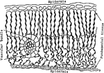
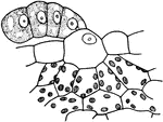
The epidermis of an oak leaf.

Plant Epidermis

The epidermis of an oak leaf.

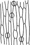
The epidermis of an iris leaf.

Plant Epidermis

The epidermis of an iris leaf.

"Cross section of a portion of leaf of Ficus elastica showing the multiple epidermis from e to a inclusive; c, cystolith; b, palisade parenchyma; d, spongy parenchyma." -Stevens, 1916

F. Elastica Epidermis

"Cross section of a portion of leaf of Ficus elastica showing the multiple epidermis from e to a inclusive;…

"A, cross section through upper half of leaf of Pyrus Japonica, showing cutinized layer of the outer wall at f, and cellulose layer at g." -Stevens, 1916

P. Japonica Epidermis

"A, cross section through upper half of leaf of Pyrus Japonica, showing cutinized layer of the outer…

A cross section through the upper half of a Russian olive leaf, showing a "cutinized layer of the outer wall at f, and cellulose layer at g." -Stevens, 1916

Russian Olive Leaf Epidermis

A cross section through the upper half of a Russian olive leaf, showing a "cutinized layer of the outer…
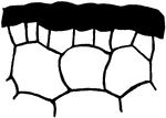
"A, portion of cross section of leaf of Avicennia growing in salty soil; outer wall of epidermis very thick." -Stevens, 1916

Avicennia Epidermis

"A, portion of cross section of leaf of Avicennia growing in salty soil; outer wall of epidermis very…
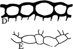
"D, upper, and E, lower epidermis of leaf of Hibiscus moscheutos." -Stevens, 1916

H. Moscheutos Epidermis

"D, upper, and E, lower epidermis of leaf of Hibiscus moscheutos." -Stevens, 1916

"Multiple epidermis of leaf of mangrove in cross section. This serves as a water reservoir, and the relatively thick walls of the inner cells reenforce the protective power of the outer layer." -Stevens, 1916

Multiple Epidermis

"Multiple epidermis of leaf of mangrove in cross section. This serves as a water reservoir, and the…
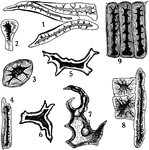
"Stone cells from different sources. 1, from coffee; 2, 3, and 4, from stem of clove; 5 and 6, from tea leaf; 7, 8, and 9, from powdered star anise seed." -Stevens, 1916

Stone Cells

"Stone cells from different sources. 1, from coffee; 2, 3, and 4, from stem of clove; 5 and 6, from…

"Diagram showing the relation of the water-carrying tissues of the leaves to those of the stem, and how the older rings of growth give up their water to the newer before this water can enter the leaves. The figures at the bottom indicate in years the age of the rings." -Stevens, 1916

Water Flow in Plants

"Diagram showing the relation of the water-carrying tissues of the leaves to those of the stem, and…
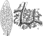
A: "Camera-lucida drawing of a bleached leaf of a Dicotyledon, showing the course of the vascular bundles, and how end free in the mesophyll." C:magnified detail. -Stevens, 1916

Dicotyledon Leaf

A: "Camera-lucida drawing of a bleached leaf of a Dicotyledon, showing the course of the vascular bundles,…
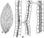
B: "Camera-lucida drawing of a bleached leaf of... a Monocotyledon, showing the anastomosis of the parallel veins by means of slender lateral branches." D: magnified detail. -Stevens, 1916

Monocotyledon Leaf

B: "Camera-lucida drawing of a bleached leaf of... a Monocotyledon, showing the anastomosis of the parallel…

"Semi-diagrammatic cross section of a leaf showing by arrows how the water passes from the tracheal elements of a vein into the border parenchyma cells, and thence into the palisade and spongy parenchyma, from which it evaporates into the intercellular spaces and passes from the leaf through the stomata. a, upper epidermis; b, lower epidermis; c, palisade parenchyma; g, spongy parenchyma; d, border parenchyma; e, tracheal elements; and the stippled cells bellow e, the phloem cells." -Stevens, 1916

Leaf Water Flow

"Semi-diagrammatic cross section of a leaf showing by arrows how the water passes from the tracheal…

"Diagram to show the path of the water as it rises to, and escapes from, the leaves." -Stevens, 1916

Leaf Water Flow

"Diagram to show the path of the water as it rises to, and escapes from, the leaves." -Stevens, 1916

"A, depressed stoma of the under side of a leaf of Amherstia nobilis." -Stevens, 1916

Depressed Stoma

"A, depressed stoma of the under side of a leaf of Amherstia nobilis." -Stevens, 1916
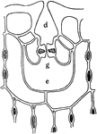
"B, depressed stoma of Hakea suaveolens. g stands beneath the guard cells; d, outer, and e inner, cavities." -Stevens, 1916

Depressed Stoma

"B, depressed stoma of Hakea suaveolens. g stands beneath the guard cells; d, outer, and e inner, cavities."…
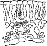
"Cross section of a portion of the blade of a leaf, showing upper epidermis at a, lower epidermis at b, palisade parenchyma at c, spongy parenchyma at d, and tracheids from the end of a vein at e." -Stevens, 1916

Leaf Epidermis

"Cross section of a portion of the blade of a leaf, showing upper epidermis at a, lower epidermis at…
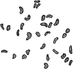
"Starch grains from the palisade cells of a Euphorbia leaf." -Stevens, 1916

Euphorbia Palisade Cells

"Starch grains from the palisade cells of a Euphorbia leaf." -Stevens, 1916
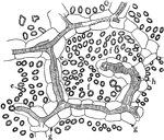
"Diagram to show the three general tissue regions of a leaf." -Stevens, 1916

Leaf Tissues

"Diagram to show the three general tissue regions of a leaf." -Stevens, 1916

"Showing intercellular spaces: f, between the palisade cells; e, in a leaf; g, border parenchyma; h, tracheal elements of the vein." -Stevens, 1916

Intercellular Spaces of Leaf

"Showing intercellular spaces: f, between the palisade cells; e, in a leaf; g, border parenchyma; h,…
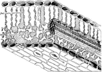
"Diagram to show the architecture of a typical leaf in the region of one of the lateral veins. The shaded parts amongst the palisade and spongy parenchyma represent intercellular spaces." -Stevens, 1916

Leaf Architecture

"Diagram to show the architecture of a typical leaf in the region of one of the lateral veins. The shaded…
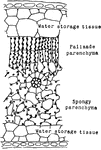
"Cross section through a portion of rubber leaf, showing the large percentage of water-storage tissues on both sides of the leaf, and the relation of the palisade and spongy parenchyma to the lateral veins." -Stevens, 1916

Rubber Leaf Cells

"Cross section through a portion of rubber leaf, showing the large percentage of water-storage tissues…
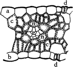
"Cross section of a portion of a leaf of Indian corn. a, upper and b, lower epidermis; c, c, palisade cells; n, border parenchyma containing starch within its chloroplasts; e, vascular bundle; d, d, stomata." -Stevens, 1916

Indian Corn Leaf Cells

"Cross section of a portion of a leaf of Indian corn. a, upper and b, lower epidermis; c, c, palisade…

"Cross section of a portion of leaf of Codonanthe, showing the water-storage tissue at f, and the chlorophyll-bearing tissues at e." -Stevens, 1916

Codonanthe Leaf Tissues

"Cross section of a portion of leaf of Codonanthe, showing the water-storage tissue at f, and the chlorophyll-bearing…
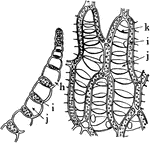
"Portion of leaf of Sphagnum, in cross section on the left, and surface view on the right. h, hole through the wall; i, chlorophyll-bearing cells; j, water-storage and water-conducting cells, with annular thickenings at k." -Stevens, 1916

Sphagnum Leaf

"Portion of leaf of Sphagnum, in cross section on the left, and surface view on the right. h, hole through…

"Cross section through a portion of leaf of Polytrichum commune. b, chains of chlorophyll-bearing cells; d, bast fibers." -Stevens, 1916

P. Commune Leaf

"Cross section through a portion of leaf of Polytrichum commune. b, chains of chlorophyll-bearing cells;…
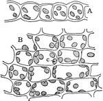
"Cross section, A, and surface view, B, of a leaf of common moss, showing chloroplasts, c." -Stevens, 1916

Moss Leaf Chloroplasts

"Cross section, A, and surface view, B, of a leaf of common moss, showing chloroplasts, c." -Stevens,…

"Showing the effect of cutting across the veins on the removal of food from the leaf. A, all of the main veins are cut across near their bases; B, the mid-vein alone has been severed. The stippled areas indicated the starch reaction with the iodine test." -Stevens, 1916

Cut Leaf Veins

"Showing the effect of cutting across the veins on the removal of food from the leaf. A, all of the…
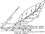
"Diagram illustrating the descent of food from the leaf into the stem, and its circulation upward and downward through the sieve tubes, and upward through the tracheal tissues." -Stevens, 1916

Leaf Food Circulation

"Diagram illustrating the descent of food from the leaf into the stem, and its circulation upward and…

"Diagram to show the relation of the food-conducting tissues of the leaf to those of the stem; and in the stem the relation of these tissues of one year to those of preceding years. The dotted line between phloem and xylem stands for the cambium. The figures at the bottom of the diagram indicate the age in years of the zones of tissues in phloem and xylem." -Stevens, 1916

Food Tissues

"Diagram to show the relation of the food-conducting tissues of the leaf to those of the stem; and in…
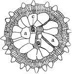
"Cross section of leaf of Mesembryanthemum Forskalii showing a large part of the leaf devoted to the storage of water. a, water-storage cells; b, chlorophyll-bearing cells; c, crystal of calcium oxalate." -Stevens, 1916

M. Forskalli Leaf

"Cross section of leaf of Mesembryanthemum Forskalii showing a large part of the leaf devoted to the…
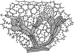
"Water-storage tracheids in the leaf of Euphorbia splendens. b, b, water-storage tracheids; d, mesophyll cells; c, branch from a milk tube." -Stevens, 1916

E. Splendens Leaf

"Water-storage tracheids in the leaf of Euphorbia splendens. b, b, water-storage tracheids; d, mesophyll…
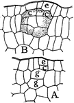
"Formation of an interior, globular, lysigenous gland of the leaf of Dictamnus fraxinella. A, g, g and c, mother cells of the gland; c, from the protoderm, and g, g, from the fundamental tissue. B, older stage where the cells have begun to form the secretion." -Stevens, 1916

D. Fraxinella Gland Formation

"Formation of an interior, globular, lysigenous gland of the leaf of Dictamnus fraxinella. A, g, g and…
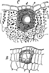
"Lysigenous gland in the leaf of Dictamnus fraxinella. B, young gland, with cells beginning to secrete oil; C, mature gland where the secreting cells have broken down and left their secretion within the cavity thus formed; o, large drop of secreted oil." -Stevens, 1916

Leaf Gland

"Lysigenous gland in the leaf of Dictamnus fraxinella. B, young gland, with cells beginning to secrete…

"Resin duct in leaf of Pinus silvestris, in cross section at A, and in longitudinal section at B; h, cavity surrounded by the secreting cells; f, f, sclerenchyma fibers surrounding and protecting the duct." -Stevens, 1916

Leaf Resin Duct

"Resin duct in leaf of Pinus silvestris, in cross section at A, and in longitudinal section at B; h,…

"Glands from Pinguicula. A, upper surface of leaf showing long-stalked gland at m, and short-stalked gland at n. B, cross section through a short-stalked gland." -Stevens, 1916

Leaf Glands

"Glands from Pinguicula. A, upper surface of leaf showing long-stalked gland at m, and short-stalked…

"Cystoliths from the leaf of Ficus carica. A, complete cystolith; B, cystolith from which the calcium carbonate has been removed for use in other parts of the plant. B is from a leaf that had fallen off in autumn." -Stevens, 1916

Cystoliths

"Cystoliths from the leaf of Ficus carica. A, complete cystolith; B, cystolith from which the calcium…

"Glands from the leaf of Ribes nigrum. A, young stage in the development of the gland where the cuticle is already being pushed up by the secretion, i. B, complete gland; k, secreting cells; h, cavity between the secreting cells and cuticle occupied by the secretion." -Stevens, 1916

Leaf Glands

"Glands from the leaf of Ribes nigrum. A, young stage in the development of the gland where the cuticle…
"Hydatode from the leaf of Phaseolus multiflorus." -Stevens, 1916

P. Multiflorus Hydatode

"Hydatode from the leaf of Phaseolus multiflorus." -Stevens, 1916

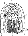
"Radial longitudinal section through a hydatode from the leaf margin of Primula sinensis. i, upper, and j, lower epidermis; h, palisade cell; e, thin-walled parenchyma, called epithem; g, intercellular space; f, guard cell of a water stoma; k, tracheal elements." -Stevens, 1916

P. Sinensus Hydatode

"Radial longitudinal section through a hydatode from the leaf margin of Primula sinensis. i, upper,…

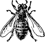
The Leaf-cutter bee, Megachile latimana.

M. Latimana

The Leaf-cutter bee, Megachile latimana.

Of the buckwheat family (Polygonaceae), the golden dock or Rumex persicarioides. The smaller image is the leaf of the swamp dock or Rumex verticillatus.

Golden Dock

Of the buckwheat family (Polygonaceae), the golden dock or Rumex persicarioides. The smaller image is…

Of the buckwheat family (Polygonaceae), the bitter dock or Rumex obtusifolius. The smaller image is the leaf of the great water dock or Rumex Britannica.

Bitter Dock

Of the buckwheat family (Polygonaceae), the bitter dock or Rumex obtusifolius. The smaller image is…