Back view of the body and outline of skeleton and covering.

Back View of the Body

Back view of the body and outline of skeleton and covering.

Lateral view of the spinal column. Labels: 1, atlas; 2, dentata 3, seventh cervical vertebra; 4, twelfth dorsal vertebra; 5, fifth lumbar vertebra; 6, first piece of sacrum; 7, last piece of sacrum; 8, coccyx; 9, a spinous process; 10, 10, intervertebral foramina.

Spinal Column

Lateral view of the spinal column. Labels: 1, atlas; 2, dentata 3, seventh cervical vertebra; 4, twelfth…
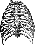
Front view of the bones of the thorax, including the ribs, sternum and vertebrae. Labels: 1, first bone of the sternum; 2, second bone of the sternum; 3, third bone or ensiform cartilage; 4, first dorsal vertebra; 5, last or twelfth dorsal vertebra; 6, first rib; 7, its head; 8, its neck; 9, its tubercle; 10, seventh or last true rib; 11, its cartilage; 12, angle of eleventh rib; 13, its body.

The Bones of the Thorax

Front view of the bones of the thorax, including the ribs, sternum and vertebrae. Labels: 1, first bone…

Anterior view of humerus (bone of the leg) of the right side. Labels: 1, shaft or diaphysis; 2, the head; 3, anatomical neck; 4, greater tuberosity; 5, lesser tuberosity; 6, the bicipital head; 3, anatomical neck; 4, greater tuberosity; 5, lesser tuberosity; 6, the bicipital groove; 7, external bicipital ridge for pectoralis major; 8, internal bicipital ridge; 9, point of insertion of deltoid muscle: 10, nutritious foramen; 11, face for head of the radius; 12, face for the ulna.

Humerus

Anterior view of humerus (bone of the leg) of the right side. Labels: 1, shaft or diaphysis; 2, the…

Anterior view of radius (bone of the arm) of the right side. Labels: 1, cylindrical head; 2, surface for lesser sigmoid cavity of the ulna; 3, neck of the radius; 4, its tubercle, for insertion of biceps muscle; 5, interosseous ridge; 5, interosseous ridge; 6, concavity for lower end of the ulna; 7, lower end of the ulna; 7, carpal surface; 8, styloid process; 9, surface for pronator quadratus muscle.

Radius

Anterior view of radius (bone of the arm) of the right side. Labels: 1, cylindrical head; 2, surface…

Anterior view of the ulna (bone of the arm) of the left side. Labels: 1, olecranon process; 2, greater sigmoid cavity; 5, external surface; 6, ridge for interosseous ligament; 7, small head for the radius; 8, carpal surface.

Ulna

Anterior view of the ulna (bone of the arm) of the left side. Labels: 1, olecranon process; 2, greater…

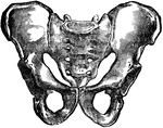
Anterior view of the male pelvis.

Male Pelvis

Anterior view of the male pelvis.

Posterior view of the femur (bone of the leg). Labels: 1, depression for round ligament; 2, the head; 3, depression for rotary muscles; 4, trochanter major; 5, trochanter minor; 6, roughness for gluteus maximus tendon; 7, 7, linea aspera; 8, surface for gastrocnemius muscle; 9, external condyle; 10, depression for anterior crucial ligament; 11, depression for posterior crucial ligament; 12, origin of internal lateral ligament.

Femur

Posterior view of the femur (bone of the leg). Labels: 1, depression for round ligament; 2, the head;…

Back view of legs, outlining bones and showing muscles.

Back View of Legs

Back view of legs, outlining bones and showing muscles.

Anterior view of the tibia (bone of the leg). Labels: 1, spinous process; 2, surface for condyles of the femur; 3, face for head of the fibula; 4, the head; 5, the tubercle; 6, 6, spine and ankle; 9, tarsal surface; 10, face for lower end of fibula.

Tibia

Anterior view of the tibia (bone of the leg). Labels: 1, spinous process; 2, surface for condyles of…

Muscle of the body, back view: The fascia is left upon the left limbs, removed from the right.

Back View of the Muscles of the Body

Muscle of the body, back view: The fascia is left upon the left limbs, removed from the right.

Muscle of the body, front view: On the right half, the superficial muscles; left half, deep-seated muscles.
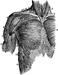
Front View of the Muscles of the Body

Muscle of the body, front view: On the right half, the superficial muscles; left half, deep-seated muscles.

Front view of the body, showing deep muscles.

Deep Muscles of the Trunk of the Body

Front view of the body, showing deep muscles.

Back view of the trunk of body, showing spinal deep muscles.

Deep Muscles of the Trunk of the Body

Back view of the trunk of body, showing spinal deep muscles.

Superior muscles of the upper front of the trunk. Labels: 1, sterno-hyoid; 2, sterno-cleido-mastoid; 3, sterno-thyroid; 4, sterno-cleido-mastoid; 5, edge of the trapezius; 6, clavicle; 7, clavicular origin of the pectoralis major; 8, deltoid; 9, fold of the pectoralis major of the anterior edge of the axilla; 10, middle of the pectoralis major; 11, crossing and interlocking of the fibers of the external oblique of one side with those of the other; 12, biceps flexor cubiti; 13, teres major; 14, serratus major anticus; 15, superior heads of external oblique interlocking with serratus major.

Muscles of the Upper Trunk

Superior muscles of the upper front of the trunk. Labels: 1, sterno-hyoid; 2, sterno-cleido-mastoid;…

Outer layer of muscles on the front of the forearm. Labels: 1, biceps flexor cubiti; 2, brachialis internus; 3, triceps; 4, pronator radii teres; 5, flexor carpi radialis; 6, palmaris longus; 7, flexor sublimus digitorum; 8, flexor carpi ulnaris; 9, palmar fascia; 0, palmaris brevis muscle; 11, abductor pollicis manur; 12, flexor brevis pollicis manus; 13, supinator longus; 14, extensor ossis metacarpi pollicis.

Muscles of the Forearm

Outer layer of muscles on the front of the forearm. Labels: 1, biceps flexor cubiti; 2, brachialis internus;…

Muscles of the front of the leg. Labels: 1, tendon of quadriceps; 2, spine of tibia; 3, tibialis anticus; 4, extensor communis digitorum; 5, extensor proprius pollicis; 6, peroneus tertius; 7, peroneus longus; 8, peroneus brevis; 9, soleus; 10, gastrocnemius; 11, extensor brevis digitorum.

Muscles of the Front of the Leg

Muscles of the front of the leg. Labels: 1, tendon of quadriceps; 2, spine of tibia; 3, tibialis anticus;…

Deep dissection of the front of the forearm and hand, showing the muscles, nerves, blood vessels, etc. Labels: 1, supinator longus (cut); 2, ulnar nerve; 3, brachialis anticus; 4, biceps; 5, musculo-spiral nerve; 6, median nerve; 7, posterior interosseous nerve; 8, pronator teres and flexor carpi radialis (cut); 9, extensor carpi radialis longior (cut); 10, brachial artery; 11, supinator brevis; 12, flexor sublimis digitorum (cut); 13, 13, radial nerve; 14, 14, flexor carpi ulnaris; 15, extensor carpi radialis brevior; 16, ulnar artery; 17, radial origin of flexor sublimis digitorum (cut); 18, flexor profundus digitorum; 19, tendon of pronator teres (cut); 20, 20, dorsal branch of ulnar nerve; 21, 21, radial artery; 22, 22, deep branch of ulnar nerve; 23, flexor longus pollicis; 24, abductor minimii digiti; 25, anterior interosseous nerve; 26, digital branches of ulnar nerve; 27, tendon of supinator longus (cut); 28, one of the lumbricales muscles (cut); 29, pronator quadratus (cut open); 31, tendon of flexor carpi radialis (cut); 33, digital branches of median nerve; 35, abductor pollicis.

Forearm and Hand

Deep dissection of the front of the forearm and hand, showing the muscles, nerves, blood vessels, etc.…
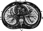
Diagram of a transverse section of the thorax. Labels: 1, anterior mediastinum; 2, internal mammary vessels; 3, triangularis sterni muscle; 4, right phrenic nerve between pleura and pericardium; 5, left phrenic nerve between the pleura and pericardium; 6, thoracic duct in posterior mediastrinum; 7, esophagus with left vagus in front and right vagus behind; 8, vena azygos major; 9, thoracic aorta giving off intercostal arteries; 10, gangliated cord of sympathetic; R.V., right ventricle; R.A., right auricle of heart in middle mediastinum; P.A., pulmonary artery; A., aorta; C., vena cava superior; V. dorsal vertebra.

The Thorax

Diagram of a transverse section of the thorax. Labels: 1, anterior mediastinum; 2, internal mammary…

A diagram of nerve tubules A nerve tube consists of a white portion which is fatty, and which protects the central portion, or axis cylinder. Surrounding these is a tubular membrane composed of epithelial cells. The nerve of the body are made up of bundles of these tubes, bound together by connective tissue and enclosed in a sheath of the same. Labels: a, axis cylinder; b, inner border of white substance; c, c, outer border of same; d, ,d, tubular membrane; B, tubular fibers; e, in natural state; f, under pressure; g, g', varicose fibers.

Nerve Tubules

A diagram of nerve tubules A nerve tube consists of a white portion which is fatty, and which protects…

Anterior view of the brain and spinal marrow. Labels: 1, 1, hemispheres of the cerebrum; 2, great middle fissure; 3, cerebellum; 4, olfactory nerves; 5, optic nerves; 6, corpora albicantia; 7, motor oculi nerves; 8, pons Varolii; 9, fourth pair of nerves; 10, lower portion of the medulla oblongata; 11, 11, medulla spinalis in its whole length; 12, 1, spinal nerves; 13, cauda equina.

Brain and Spinal Cord

Anterior view of the brain and spinal marrow. Labels: 1, 1, hemispheres of the cerebrum; 2, great middle…
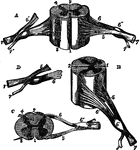
Different views of a portion of the spinal cord from the cervical region, with roots of the nerves slightly enlarged. Labels: In A, the anterior surface of the specimen is shown, the anterior nerve root of its right side being divided: in B, a view of the right side is given; in C, the upper surface is shown; in E, the nerve roots and ganglion are shown from below; 1, the anterior median fissure; 2, posterior median fissure; 3, anterior lateral depression, over which the anterior nerve roots are seen to spread; 4, posterior lateral groove, into which the posterior roots are seen to sink; 5, anterior roots passing the ganglion; 5', in A, the anterior root divided; 6, the posterior roots, the fibers of which pass into the ganglion 6'; 7, the united or compound nerve; 7', the posterior primary branch, seen in A and D to be derived in part from the anterior and in part from the posterior root.

Different View of the Spinal Cord

Different views of a portion of the spinal cord from the cervical region, with roots of the nerves slightly…

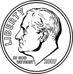
An illustration of the portrait side of a U.S. Dime.

Portrait on a Dime

An illustration of the portrait side of a U.S. Dime.

An illustration of the portrait side of a U.S. Dollar Coin.

Portrait on a Dollar

An illustration of the portrait side of a U.S. Dollar Coin.

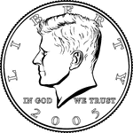
An illustration of the portrait side of a U.S. Half Dollar.

Portrait on a Half Dollar

An illustration of the portrait side of a U.S. Half Dollar.

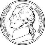
An illustration of the portrait side of a U.S. Nickel.

Portrait on a Nickel

An illustration of the portrait side of a U.S. Nickel.

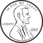
An illustration of the portrait side of a U.S. Penny.

Portrait on a Penny

An illustration of the portrait side of a U.S. Penny.

An illustration of the portrait side of a U.S. Quarter.

Portrait on a Quarter

An illustration of the portrait side of a U.S. Quarter.

Half of the teeth of the upper jaw. <em>a,a,</em> are the two front cutting teeth. <em>d,d,d</em> are the three large back teeth. c,c are two smaller grinders. At <em>b</em> is what is commonly called an "eye-tooth."

Teeth of the Upper Jaw

Half of the teeth of the upper jaw. a,a, are the two front cutting teeth. d,d,d are…
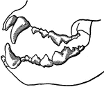
Teeth of a carnivorous animal that lives on flesh alone. The front teeth are designed for tearing, while the back teeth have sharp edges for cutting.

Teeth of a Carnivorous Animal

Teeth of a carnivorous animal that lives on flesh alone. The front teeth are designed for tearing, while…

Teeth of an herbivore, showing the rough surface of some of these teeth. Herbivores have no tearing teeth. Instead they have two kinds of teeth, cutting teeth in the front and grinding teeth in the back. This figure shows a peculiar arrangement of the enamel, which admirably fits them to grind up the fibers of the grass in the back grinding teeth of an herbivore. The enamel is not only on the outside as it is in human teeth, rather there are ridges on it standing up in the middle of each tooth.

Teeth of an Herbivore

Teeth of an herbivore, showing the rough surface of some of these teeth. Herbivores have no tearing…

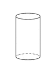
A flashcard featuring an illustration of a Cylinder

Flashcard of a Cylinder

A flashcard featuring an illustration of a Cylinder

A representation of the heart as it really appears showing the front view. At <em>a</em> is the right auricle that receives blood from all pats of the body by two large veins h and i. At <em>b</em> is the right ventricle, which receives the blood from the auricle and sends it to the lungs by the pulmonary artery <em>f</em>. At <em>c</em> is the left auricle, which receives the blood from the lungs by the pulmonary veins <em>g, g, g</em>. At <em>d</em> is the left ventricle which receives blood from the auricle and forces it all over the body through the aorta <em>e</em>.

Heart, Front View of

A representation of the heart as it really appears showing the front view. At a is the right…
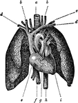
The heart, showing its relative position to the lungs. The heart is almost wholly covered up by the lungs and it is encased in a sack or bag (the pericardium), and around this there is considerable padding. The lungs are represented as drawn apart, so that a full view of the heart with its arteries and veins can be seen, and the sac of the heart and packing material is also removed. At <em>a</em> is the trachea or windpipe; on each side are the two arteries that go to the head; <em>c</em> is the artery that goes to the arm; <em>b, b</em> are the veins coming from the head, and <em>d, d</em> the veins from the arms, all emptying into a large vein that goes to the right auricle of the heart, <em>e</em>; <em>f</em> is the large vein that brings the blood from below to this auricle; <em>g</em> is the right ventricle, <em>i</em> the left, and <em>h</em> is the aorta as it goes down from the heart.

Heart and Lungs

The heart, showing its relative position to the lungs. The heart is almost wholly covered up by the…
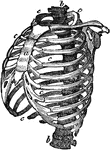
Structure of the chest, showing the framework of the bones which are connected together chiefly by muscles. It is important to understand the structure of the chest in order to understand how the movements of the chest are made in inspiration and expiration. The spinal column <em>b,b</em> is the grand pillar that supports this barrel-shaped framework. The ribs <em>c, c, c</em> are fastened very strongly by ligaments to the spinal column. They are 2 in number, 12 on each side and extend round towards the breast bone (sternum) <em>a</em> in front.

Structure of the Chest

Structure of the chest, showing the framework of the bones which are connected together chiefly by muscles.…

The diaphragm, which is the principal muscle that act sin breathing. Here you have the cavity of the chest, <em>Cc</em>, laid open, the ribs being cut away in front, and the heart and lungs taken out; <em>DD</em> is the diaphragm. It is fastened to the spinal column behind, to the breast-bone in front, and to the lower ribs all around its side. It is not flat, but arched upward.

The Diaphragm

The diaphragm, which is the principal muscle that act sin breathing. Here you have the cavity of the…

Diaphragm of the diaphragm and it's placement in the chest. Let <em>a</em> represent the spinal column, <em>b</em> the front wall of the chest, <em>Cc</em> the cavity of the chest, and <em>Ca</em> the cavity of the abdomen. At <em>d</em> is represented the diaphragm. As you can see from the figure, as the fibers of the diaphragm are shortened the space in the chest increases which is what takes place during inspiration. On the contrary, during expiration the diaphragm is pushed upward as shown in this figure so the room in the chest is lessened and air is forced out.

The Diaphragm

Diaphragm of the diaphragm and it's placement in the chest. Let a represent the spinal column,…
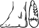
On the left is a side view of a Chinese lady's foot that has been bound while it was growing to stunt it's growth. On the right is a view of the sole of the same foot. All the toes but the great one are turned in under the foot.

Foot of Chinese Woman

On the left is a side view of a Chinese lady's foot that has been bound while it was growing to stunt…
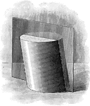
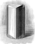
Illustration of a triangular prism inscribed in a cylinder.

Prism inscribed in Cylinder

Illustration of a triangular prism inscribed in a cylinder.

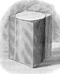
Illustration of a prism circumscribed about a cylinder.

Prism Circumscribed About A Cylinder

Illustration of a prism circumscribed about a cylinder.

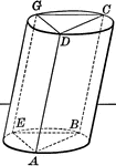
Illustration of a plane passing through a cylinder. Every section of a cylinder made by a plane passing through an element is a parallelogram.

Plane Passing Through A Cylinder

Illustration of a plane passing through a cylinder. Every section of a cylinder made by a plane passing…
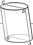
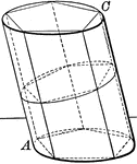
Illustration of a plane passing through a cylinder. Every section of a cylinder made by a plane passing through an element is a parallelogram.

Plane Passing Through A Cylinder

Illustration of a plane passing through a cylinder. Every section of a cylinder made by a plane passing…
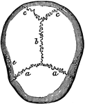
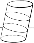
Illustration of planes passing through a cylinder.

Planes Passing Through A Cylinder

Illustration of planes passing through a cylinder.

Illustration of a triangular prism inscribed in a cylinder.

Prism inscribed in Cylinder

Illustration of a triangular prism inscribed in a cylinder.

Illustration of a pentagonal prism inscribed in a cylinder.

Prism inscribed in Cylinder

Illustration of a pentagonal prism inscribed in a cylinder.

Illustration of a cylinder of revolution.

Cylinder of Revolution

Illustration of a cylinder of revolution.

Illustration of an isosceles spherical triangle.

Solids of Revolution

Illustration of an isosceles spherical triangle.

The bones of the top of the head are fastened together by what are called sutures which are locked together by little teeth of bone that shoot by each other, as shown in the figure. <em>b</em> is the suture at the top of the head between the two parietal bones; <em>a a</em> is that between the two parietal bones and the frontal bone in front; and <em>c c </em> is that between the parietal bones and the bone at the back part of the cranium. These bones are not completely formed in the newborn infant.

Cranial Sutures

The bones of the top of the head are fastened together by what are called sutures which are locked together…

Two views of vertebra. 24 vertebrae make up the spinal column. On the left figure, <em>a</em> is the body of the bone; <em>b</em> is the hole through the vertebra which forms its part of the canal for the spinal marrow; and <em>c</em> is the spinous process. On the right is a side view of the same vertebra.

Two Views of a Vertebra

Two views of vertebra. 24 vertebrae make up the spinal column. On the left figure, a is the…

View of the entire spinal column. The bodies of the vertebrae are in the front with the spinous processes on the other side. Between the row of the bodies of the vertebrae and the row of spinous processes you can see the canal in which lies the spinal marrow.

The Spinal Column

View of the entire spinal column. The bodies of the vertebrae are in the front with the spinous processes…

The vertebra of a fish, which is very different from that of a human. It has but two processes, <em> f f </em>. In humans there is a single short spinous process behind, while the vertebra is round in front. But in the fish there are two quite long spinous processes, one in front and the other in the rear (which is above and below, respectively, when the fish is in water).

Vertebra of a Fish

The vertebra of a fish, which is very different from that of a human. It has but two processes,

Rear view of the thigh bone.

Rear View of the Thigh Bone

Rear view of the thigh bone.

Bones of the foot, side view. In this figure the bones of the tarsus extend from the heel to <em>a</em>; the metatarsal bones are at <em>b</em>; and the bones of the toes are at <em>c</em>. In every movement of the foot there is some little motion between all these bones, and it is this that gives ease and grace to its motions.

Side View of the Bone of the Foot

Bones of the foot, side view. In this figure the bones of the tarsus extend from the heel to a;…
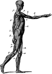
Side view of the muscles of the body, showing those that lie directly under the skin. Other deeper muscles are not visible. Muscles vary widely in their shape and size depending on their function.

Side View of the Muscles of the Body

Side view of the muscles of the body, showing those that lie directly under the skin. Other deeper muscles…

The lids of the eye close in such a way as to leave a three-cornered canal between them and the surface of the eyeball. Labels: b, the surface of the front of the eye; a, the edges of the two lids.

Eye Lid

The lids of the eye close in such a way as to leave a three-cornered canal between them and the surface…
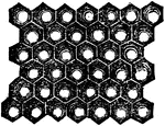
A magnified view of a small portion of the surface of an insect's compound eye which is composed of little eye-tubes.

Eye of an Insect

A magnified view of a small portion of the surface of an insect's compound eye which is composed of…

Hard Times Token (unknown) US coin. Obverse has an image of the front of the Merchants Exchange surrounded by the inscription - MERCHANTS EXCHANGE WALL ST N YORK BUILT 1827 BURNT 1835. Reverse shows the center inscription - NOT ONE CENT FOR TRIBUTE surrounded by a wreath with 9 berries - 3 outside and 6 inside the wreath with a surrounding inscription - MILLIONS FOR DEFENCE.

Copper Hard Times Token, ND

Hard Times Token (unknown) US coin. Obverse has an image of the front of the Merchants Exchange surrounded…

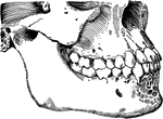
Front and side views of the teeth and jaws.

Teeth and Jaws

Front and side views of the teeth and jaws.

Projection of a round bar.

Projection of Round Bar

Projection of a round bar.

Projection of a cylinder with a circular hole.

Projection of Cylinder With Circular Hole

Projection of a cylinder with a circular hole.